Download
1 / 59
610 likes | 1.33k Vues
Nasal Cavity and Pterygopalatine Fossa. R. Shane Tubbs, MS, PA-C, PhD. columella. Five major cartilages. Piriform aperture Anterior nasal spine Nasal septum Nasal bones. Nasal Cavity: Borders. Roof: frontal, ethmoid (cribriform), sphenoid, nasal bones

E N D
Nasal Cavity and Pterygopalatine Fossa R. Shane Tubbs, MS, PA-C, PhD
columella • Five major cartilages
Piriform aperture • Anterior nasal spine • Nasal septum • Nasal bones
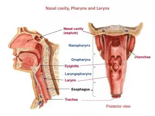
Nasal Cavity: Borders • Roof: frontal, ethmoid (cribriform), sphenoid, nasal bones • Floor: maxillary and palatine bones • Medial: nasal septum • Lateral: nasal conchae, lacrimal, maxillary, palatine bones
Nasal Septum • Perpendicular plate • Septal cartilage • Vomer • Medial crus of > alar cartilage • Nasal crests of maxillary, palatine, and sphenoid bones • Nasal spine of frontal bone vomer
Vomeronasal Cartilage • Along inferior border of septal cartilage • Rudimentary in man • Vomeronasal nerve of Jacobson in lower animals- pheromones
ethmoid maxillary inferior concha sphenopalatine foramen
bulla uncinate process
Features • Bulla (bubble) • Nasofrontal duct • Uncinate process • Semilunar hiatus
Valve of Hasner (Czech Ophthalmologist 1819-1892) • Iatrogenic closure
Features • Vestibule: skin/vibrissae, sweat and sebaceous glands • Upper 1/3 • Lower 2/3 • Limen (entrance) nasi (lateral nasal cartilage) • Agger (mound) nasi (ethmoid air cells)
Olfactory Nerves • ~ 20 pairs • Most commonly injured cranial nerve • CSF rhinorrhea • Do not regenerate in elderly
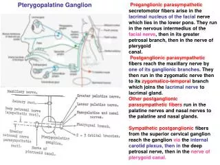
Pterygopalatine ganglion Vidian anterior ethmoidal greater palatine nasopalatine
Arterial Supply • Sphenopalatine • Anterior ethmoidal • Posterior ethmoidal • Greater palatine • Superior labial and lateral nasal branch of facial
Nasal Veins/Lymphatics • Veins: Drain via sphenopalatine foramen into pterygoid plexus and some via ethmoidal foramina to superior ophthalmic vein • Lymphatics: Majority join pharyngeal plexus and thus drain into retropharyngeal nodes
Paranasal Air Sinuses • Function • Named for the bones they occupy • Paired • Surrounded by diploic space of contiguous bones
Frontal Sinus • Frontonasal duct- semilunar hiatus • Innervation: supraorbital n. • Variation • Acromegaly • Eskimos • Related to anterior cranial fossa • Tubbs et al. J Neurosurgery, 2002
Ethmoid Sinus (3-18 pairs) Named on the basis of their openings anterior: semilunar hiatus middle: ethmoidal bulla or directly into middle meatus posterior: superior meatus Innervation: anterior and posterior ethmoidal nerves and branches of pterygopalatine ganglion
Sphenoid Sinus • Sphenoethmoidal recess • Most variable cavity in the body! • 15% of all cases of sinusitis • Ostium is 1.5 cm superior to its floor • Innervation: Posterior ethmoidal nerve and branches of pterygopalatine ganglion • Related to middle cranial fossa
Maxillary Sinus • Maxillary: semilunar hiatus • Innervation: ant, middle, posterior superior alveolar nerves, infraorbital (V2) • Most commonly infected sinus • Drains superiorly as does sphenoid sinus
Pterygopalatine Fossa • “A pyramidal space inferior to the apex of the orbit and lateral to the nasal cavity” • ~ 2 x 1 cm • Arteries: post sup alveolar, descending palatine, pterygoid canal, pharyngeal, sphenopalatine • Maxillary nerve • Nerve of pterygoid canal (Vidian) • Pterygopalatine ganglion (posterior to middle nasal concha) • Pterygopalatine nerves
Four canals: Vidian, vomerovaginal, palatovaginal, greater palatine canal • Two foramina: rotundum, sphenopalatine • Two fissures: inferior orbital, pterygomaxillary
Pterygopalatine Fossa • Lateral: pterygomaxillary fissure • Medial: perpendicular plate of palatine with sphenopalatine foramen • Posterior: Pterygoid process with Vidian canal, rotundum • Anterior: maxillae with inferior orbital fissure, posterior superior alveolar foramen (lateral) • Roof: > wing sphenoid, superior orbital fissure • Inferior: pyramidal process, palatine canal (oral cavity)
Pterygopalatine Ganglion • Parasympathetic root • Sympathetic root • Sensory root
Distribution of Pterygopalatine Ganglion • Sphenoid sinus (pharyngeal branch) • Posterior ethmoid cells • Nose • Hard and soft palate • Inner gingivae of maxillary teeth • Palatine tonsil • Choana • Uppermost pharynx • Orbit