Download
1 / 39
570 likes | 1.84k Vues
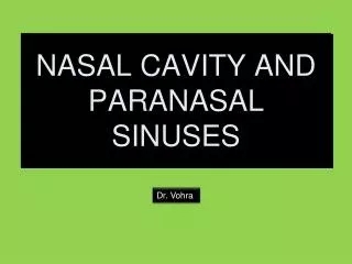
NASAL CAVITY AND PARANASAL SINUSES. Dr. Vohra. THE NOSE. THE NOSE. The external nose has a free tip ( apex ) and a root ( bridge ). The external orifices are the two nostrils ( nares ). Each nostril is bounded medially by the nasal septum. THE NOSE.

E N D
NASAL CAVITY AND PARANASAL SINUSES Dr. Vohra
THE NOSE • The external nose has a free tip (apex) and a root (bridge). • The external orifices are the two nostrils (nares). • Each nostril is bounded medially by the nasal septum. Dr. Vohra
THE NOSE The nostrils are bounded laterally by the alla. Dr. Vohra
NOSE The framework of the nose is made: • Above by : • The nasal bones; • The frontal processes of the maxillae; • The nasal part of the frontal bone • Below by hyaline carilages: • Upper nasal cartilages • Lower nasal cartilages • Septalcatilage Dr. Vohra
Blood Supply of the External Nose The skin of the external nose is supplied by branches of the ophthalmic and the maxillary arteries. The skin of the ala and the lower part of the septum are supplied by branches from the facial artery. Nerve Supply of the External Nose The infratrochlear and external nasal branches of the ophthalmic nerve (CN V) and the infraorbital branch of the maxillary nerve
NASAL CAVITY BOUNDARIES NERVE SUPPLY BLOOD SUPPLY LYMPH DRAINAGE
BOUNDARIES • From the nostrils in front • to the choanae behind • Divided into right and left half by the nasal septum • Each half has: • Floor • Roof • Lateral wall • Medial wall Dr. Vohra
Boundaries of the Nasal Cavity • The floor (this is the upper surface of the hard palate) is made from: • The palatine process of the maxilla and • The horizontal process of the palatine bone • The roof is narrow and it is formed, from behind forward, by: • The body of the sphenoid • The cribriform plate of the ethmoid • The frontal bone • The nasal bone • The nasal cartilages Dr. Vohra
The lateral wall of the nose • The lateral wall has three projections called the: • Superior concha • Middleconcha • Inferiorconcha • The area below each concha is called a meatus. Dr. Vohra
The lateral wall of the nose • The area below each concha is called a meatus. • Superior meatus • Middlemeatus • Inferior meatus Dr. Vohra
Openings in the Superior Meatus • The superiormeatus lies below and lateralto the superior concha. • It receives the openings of the posterior ethmoidal air sinuses • The sphenoethmoidal recess lies anterior to the body of the sphenoid bone. • It receives the opening of the sphenoidal air sinus. Dr. Vohra
Openings in the Middle Meatus • The middle meatus lies below and lateral to the middle concha. • It has on its lateral wall a rounded prominence, the bulla ethoidalis. • Bulla ethmoidalisis caused by the bulging of the underlying middle ethmoidal air sinuses, which open on its upper border. • Hiatus semilunarisis a curved cleft below the bulla. • It leads into a funnel-shaped channel called theinfundibulum • The frontal sinusopens into and is continuous with the infundibulum. Dr. Vohra
Openings in the Middle Meatus • The maxillary sinus opens in the middle meatus via hiatus semilunaris. • The anterior ethmoidal sinusesalso open in the infundibulum. Dr. Vohra
Structures related to the middle meatus • The middle meatus is coninuousanteriorly with a depression called the antrum. • The antrum is limited above by a ridge called aggernasi. • Below and in front of the antrum is the vestibule. • It is lined by modified skin and has short hairs called vibrissae. Dr. Vohra
Openings in the Inferior Meatus • The inferior meatus lies below and lateral to the inferior concha. • It receives the opening of thenasolacrimal duct. • The opening of the nasolacrimal duct is garded by a fold of mucosa, forming an imperfect valve. Dr. Vohra
Medial wall of the nose • The medial wall (the nasal septum) is an osteocartilaginous partition, covered by mucous membrane. • It is formed by: • the vertical (perpendicular) plate of the ethmoid bone, • the vomer and • the septal cartilage. Dr. Vohra
MUCOUS MEMBRANE • It lines all the nasal cavity, with the exception of the vestibules (lined with modified skin). • Two types of mucous membrane: • olfactory • respiratory Dr. Vohra
OLFACTORY MUCOSA • On the lateral wall, it lines the upper surface of the superior concha and the spheno-ethmoidal recess. • On the medial wall, it lines the superior part of the nasal septum. Dr. Vohra
RESPIRATORY MUCOSA • It lines the lower part of the nasal cavity. • It functions to moisten, clean and warm the inspired air. • The air is moistened by the secretion of numerous serous glands. • The air is warmed by a submucous venous plexus. Dr. Vohra
NERVE SUPPLY TO THE NASAL CAVITY • The olfactory nerves arise from the central axons of the olfactory nerve cells. • They ascend through the cribriform plateto reach the olfactory bulbs. • The nerves of ordinary sensation are branches of the ophthalmic division (CnV1) and the maxillary division (CnV2) of the trigeminal nerve Dr. Vohra
ARTERIAL BLOOD SUPPLY • It comes mainly from branches of the maxillary artery. • The most important branch is the sphenopalatine artery. • This artery anastomoses with the septal branch of the superior labial artery in the region of the vestibule. • This is a common site of bleeding from the nose (epistaxis). Dr. Vohra
LYMPH DRAINAGE • The vestibule of the nasal cavity is drained into the submandibular lymph nodes. • The reminder of the nasal cavity is drained into the upper deep cervical lymph nodes. Dr. Vohra
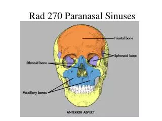
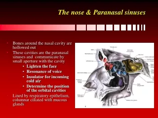
PARANASAL SINUSES MAXILLARY FRONTAL SPHENOIDAL ETHMOIDAL
DEFINITION • They are cavities inside the: • Maxilla • Frontal bone • Sphenoid bone • Ethmoid bone • They are: • Lined with mucoperiosteum; • Filled with air; • Communicate with the nasal cavity. Dr. Vohra
Functions of the Paranasal Sinuses • Reduce the weight of the skull. • Resonators of the voice. Dr. Vohra
Drainage of Mucus and Function of Paranasal Sinuses • The mucus produced by the mucosus membrane is drained into the nose by the ciliary action of the columnar cells. • It is also helped by siphon action created during blowing of the nose. • When the apertures of the sinuses are blocked or when they are filled with fluid, the quality of the voice becomes markedly changed. Dr. Vohra
MAXILLARY SINUS • It is located within the body of the maxilla behind the skin of the cheek. • It has a pyramidal form: • The roots of the: • First premolar tooth • Second premolar tooth • Third molar tooth • Canine tooth (sometimes) • - Project into the maxillary sinus • Tooth extraction can produce a fistula. • Tooth infection can produce sinusitis. Dr. Vohra
MAXILLARY SINUS The maxillary sinus opens in the middle meatus through the semilunar hiatus. Dr. Vohra
NERVE SUPPLY MAXILLARY SINUS • The mucous membrane of the maxillary sinus is supplied by the: • Superior alveolar and the • Infraorbitalnerves. Dr. Vohra
FRONTAL SINUS • The frontal sinuses (two) present within the frontal bone. • They are separated by a bony septum (frequently deviated to one side). • Each sinus is roughly triangular. • It extends: • Upward above the medial end of the eyebrow • Backward into the medial part of the roof of the orbit. • Each frontal sinus opens into the middle meatus Dr. Vohra
SPHENOIDAL SINUSES • They are two in number • They lie within the body of the sphenoid bone. • Each sinus opens into the sphenoethmoidal recess above the superior concha. • The mucous membrane is supplied by the posterior ethmoidal nerves. Dr. Vohra
ETHMOIDAL SINUSES • They are contained within the ethmoid bone. • Only a thin layer of bone separates these sinuses from the orbit. • Infection can readily spread from the ethmoidal sinuses into the orbit. Dr. Vohra
ETHMOIDAL SINUSES • They are divided into three groups: anterior, middle and posterior. • The anterior groupdrains in the infundibulum. • The middle groupdrains in the middle meatus (on or above bulla ethmoidalis). • The posterior groupdrains in the superior meatus. The mucous membrane of the ethmoidal sinuses is supplied by the anterior and posterior ethmoidal nerves. Dr. Vohra
Paranasal Sinuses and Their Site of Drainage Into the Nose Dr. Vohra
INFECTION OF THE NASAL CAVITY (RHINITIS) • Infection of the nasal cavity may spread to: • The paranasal sinuses; • Via the nasopharynx to the auditory tube and the middle ear; • To the anterior cranial fossa (via the cribriform plate) – and to produce meningitis.
NOSE BLEEDING (EPISTAXIS) • The most common cause is nose picking. • May be arterial or venous in origin. • Often occurs from the anteroinferior portion of the septum and involve the septal branches of the sphenopalatine and facial vessels.
INFECTION OF PARANASAL SINUSES (SINUSITIS) • A common complication of nasal infection. • Rarely, it could be a complication of apical dental abscess (for the maxillary sinus).