Download
1 / 20
210 likes | 302 Vues
Explore microscopic structures of the upper respiratory tract focusing on Nasal Cavity, Paranasal Sinuses, and Larynx. Learn about the vestibule, mucosa, septum, and clinical applications. Detailed content on epithelium, lamina propria, glands, and more.

E N D
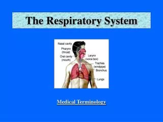
Histology of the Upper Respiratory Tract(Nasal cavity, Paranasal sinuses and Larynx) RESPIRATORY SYSTEM ( I )
Objectives: By the end of this lecture the student should be able to describe the microscopic structures of: • Vestibule of the nasal cavity. • Respiratory mucosa of the nasal cavity. • Nasal septum. • Olfactory mucosa of the nasal cavity. • Mucosa of the paranasal sinuses. • Larynx.
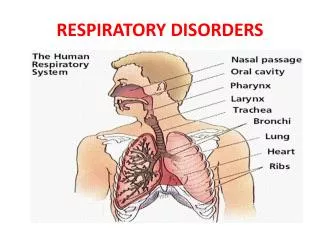
RESPIRATORYSYSTEM • Conducting portion : 1- Nasal cavity. 2- Nasopharynx. 3- Larynx. 4- Trachea. 5- Primary bronchi (extrapulmonary bronchi). 6- Intrapulmonary bronchi: - 2ry bronchi (lobar bronchi). - 3ry bronchi (segmental bronchi). 7- Primary bronchioles (preterminal bronchioles). 8- Terminal bronchioles. • Respiratory portion: 1- Respiratory bronchioles. 2- Alveolar ducts . 3- Alveolar sacs. 4- Pulmonary alveoli.
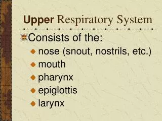
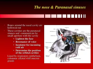
NASAL CAVITY (N.C.) • Anterior portion of N.C.: Vestibule. • Posterior portion of N.C.: a- Respiratory region. b- Olfactory region. N.B. The nasal septum divides the nasal cavity into two halves (right and left).
VESTIBULE OF N.C. Lining: is lined with thin skin. 1- Epidermis: (Keratinized stratified Squamous epithelium). 2- Dermis. Contents: 1- Vibrissae: stiff hairs. 2- Sebaceous glands. 3- Sweat glands. Wall: 1- Hyaline cartilage. 2- Cancellous (spongy) bone.
RESPIRATORY REGION (AREA) OF NASAL CAVITY MUCOSA (MUCOUS MEMBRANE): (A) Epithelium: Pseudo-stratified ciliated columnar epithelium with goblet cells (Respiratory epithelium). (B) Lamina propria ( Sub-epithelial C.T.): contains: 1- Large arterial plexuses & venous sinuses (Highly vascularized C.T.) 3- Many seromucous glands (acini). 4- Abundant lymphoid elements: Including occasional lymphoid nodules, plasma cells & mast cells.
PARANASAL SINUSES Lining: 1- Respiratory epith. (Mention…….) 2- Lamina propria. CLINICAL APPLICATION: Sinusitis.
OLFACTORY REGION (AREA) OF NASAL CAVITY(OLFACTORY MUCOSA) Site: 1-Roof of nasal cavity. 2-Upper part of nasal septum. 3-over superior concha. Structure: • Olfactory epithelium: Pseudo-stratified columnar epithelium. 1- Olfactory cells (olfactory nerve cells) 2- Sustentacular (supporting) cells. 3- Basal cells: Pyramidal in shape, basal in position and act as stem cells. (B) Lamina propria: contains: 1- Highly (richly) vascularized loose to dense C.T. 2- Contents: a) Bowman’s glands ( olfactory glands) : are serous acini. b) Bundles of unmyelinated nerve fibers: Are axons of olfactory nerve cells + Schwann-like cells (glial cells). c) Rich vascular plexus. d) Numerous lymphoid elements.
OLFACTORY EPITHELIUM 1- Olfactory cells: Are bipolar neurons Dendrite has olfactory vesicle that has nonmotile cilia. Axons are unmyelinated with Schwann-like cells. Axons will collect in the lamina propria to form bundles of nerve fibers. Bundles will collect to form the olfactory nerve. 2- Sustentacular (supporting) cells: Are columnar cells. Function: Physical support and nourishment for olfactory cells.
LARYNX • Mucosa (Mucous membrane ): 1- Epithelium. 2- Lamina propria. (B)Cartilages. (C) Extrinsic and intrinsic muscles: all are skeletal. (D) Ligaments.
LARYNX • Mucosa: 1- Epithelium: (2 types) a- Respiratory epithelium: Pseudostratified ciliated columnar epithelium with goblet cells. b- Non keratinized stratified squamous epithelium: In: -Vocal folds. - Superior surface of epiglottis 2- Lamina propria.
LARYNX • Mucosa (cont.): There are 2 pairs of shelf-like mucosal folds: 1- Vestibular folds: Are immovable. L/M: a- Respiratory epithelium. b- Lamina propria: Loose C.T. with seromucous glands lymphoid elements & adipose cells. 2- VOCAL FOLDS (CORDS): have: a- Epithelium: non keratinized stratified squamous. b- Lamina propria: C.T. containing bundles of elastic fibers and skeletal muscle . N.B. No lymphoid nodules, No seromucous glands.
(B)Cartilages: 1- Hyaline cartilages: e.g. Thyroid cartilage. 2- Elastic cartilages: Epiglottis. (C) Muscles:all are skeletal. (D) Ligaments.
RESPIRATORY EPITHELIUM Pseudo-stratified ciliated columnar epithelium with goblet cells. Main Types of cells ( all touch the basement membrane) 1- Ciliated columnar cells. 2-Goblet cells. 3-Basal cells: are stem cells. 4- DNES cells: e.g. serotonin.
RESPIRATORY SYSTEM (II) Histology of the Lower Respiratory Tract (Trachea, Bronchi, Bronchioles) & the Lung
TRACHEA The wall of trachea is formed of: • Mucosa. • Submucosa. • Adventitia.
MUCOSA OF TRACHEA • Epithelium: Respiratory epithelium • Lamina propria. (3) Elastic lamina: It is formed of elastic fibers. It separates lamina propria from submucosa.
SUBMUCOSA OF TRACHEA Contents: 1- C.T. 2- Numerous mucous & seromucous glands. 3- Lymphoid elements.
ADVENTITIA OF TRACHEA Contents: 1- Fibroelastic C.T. 2- C-shaped rings (12-16) of hyaline cartilage. Trachealis muscle (bundle of smooth muscle fibers) connects the 2 ends of each C-shaped (incomplete) rings of cartilage.