Download
1 / 16
180 likes | 239 Vues
Discover the anatomy of the nose and paranasal sinuses, their functions, development, and relation to adjacent structures. Learn about the maxillary, ethmoidal, and sphenoidal sinuses, blood supply, nerve and lymph drainage, and potential complications.

E N D
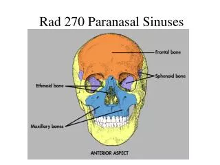
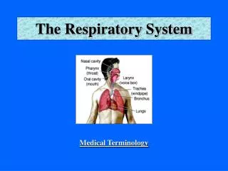
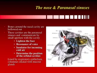
The nose & Paranasal sinuses • Bones around the nasal cavity are hollowed out • These cavities are the paranasal sinuses and communicate by small aperture with the cavity • Lighten the face • Resonance of voice • Insulator for incoming cold air • Determine the position of the orbital cavities • Lined by respiratory epithelium, columnar ciliated with mucous glands
The nose & Paranasal sinuses • The maxillary sinus: • It is pyramidal in shape with • The base at the lateral wall of the nose • The apex the zygomatic process of the maxilla. • The roof is the floor of the orbit • The floor is the alveolar process of the maxilla
The nose & Paranasal sinuses • Begins developing about the 4th month of intra-uterine life and continue to grow till the 3rd decade • It varies in size and is the largest of all the paranasal sinuses
The nose & Paranasal sinuses • It lies at a lower level than the floor of the nose. • The ostium is high up on its nasal wall, 2 – 4 mm in diameter at the posterior end of hiatus semilunaris in the middle meatus.
The nose & Paranasal sinuses • The infra-orbital nerve lie in the roof, its branches to the teeth descend along the anterolateral wall, leadind to facial and dental pain when the sinus is diseased • The upper teeth are in close relation to the floor, 1st and 2nd molar teeth • Disease of the sinus as a result of infected teeth
The nose & Paranasal sinuses maxillary sinus: • Blood supply: • Branches from the facial, maxillary, infraorbital and greater palatine arteries • Nerve supply: • supplied by branches from the superior alveolar nerves and the greater palatine nerve. • Lymph drainage: • drain into the submandibular lymph nodes
The nose & Paranasal sinuses • Ethmoidal sinus: • lies between the orbit and the nose in the ethmoidal labyrinth. • It is not a single cavity (8-10 thin-walled intercommunicating cavities) • It is divided by bony septa into three ethmoidal air cells, anterior, middle and posterior, each with it is own ostium, draining into the middle and superior meatus.
The nose & Paranasal sinuses • The lateral wall is the medial surface of the orbit • The medial wall is the lateral surface of the nasal cavity with the superior and middle conchae projecting from it
The nose & Paranasal sinuses • Above is the anterior cranial fossa and the frontal lobe • Anteriorly, is the frontal sinus • Posteriorly, the sphenoid bone • Inferiorly, the nose • Separated from these surrounding structures by thin wall • Infection (ethmoiditis) spread rapidly • is the commonest cause of orbital cellulitis • Meningitis, subdural and cerebral abscesses and cavernous sinus thrombosis
The nose & Paranasal sinuses Ethmoidal sinus: • Blood supply: • from both internal and external carotid arteries via the supraorbital, anterior and posterior ethmoidal and sphenopalatine. • Nerve supply: • from both maxillary and ophthalmic nerves, supra-orbital, anterior & posterior ethmoidal, orbital and lateral posterior nasal. Referred pain ??? • Lymph drainage: • submandibular and retropharyngeal nodes.
The nose & Paranasal sinuses • Sphenoidal sinus: • A large cavity situated in the body of the sphenoid bone • Divided by a septum that bend to one side leading to right and left halves, that vary greatly in size. • It lies beneath the pituitary fossa, extend into the basiocciput, greater wing of sphenoid and the pterygoid process. • Begins to develop at the 5th month of I-U life as a recess in the nasal bone and extend into the sphenoid bone at the age of 7
The nose & Paranasal sinuses • The roof is related to the pituitary fossa and the optic nerve – sudden loss of vision in case of sinus infection • Anteriorly, the ethmoid air cells • Lateral, the cavernous sinus containing the internal carotid artery and Abducent nerve – Stabismus and Diplopia • The floor is the nose
The nose & Paranasal sinuses • The ostium is in the anterior wall of the sinus and open into the sphenoethmoidal recess, above superior concha. • Blood supply: • posterior ethmoidal & sphenopalatine from maxillary • Nerve supply: • posterior ethmoidal • orbital branch of pterygopalatine ganglion • ophthalmic and maxillary nerve
The nose & Paranasal sinuses • Frontal sinus: • the only sinuses not present at birth, appear after the second year. • The two sinuses are unequal in extent. They are related to the anterior cranial fossa and the orbit • It present a posterosuperior wall, anterior wall and floor • Posterosuperior wall is thin and separate the sinus from the meninges and the frontal lobe of the brain • The anterior wall form the forehead • The floor separate the sinus fron the orbit, nose and anterior ethmoid sinus
The nose & Paranasal sinuses • The ostium open into the semilunar hiatus, with the other openings: • Infection travel from one sinus to the other • Deviation of the nasal septum might aggravate the infection • Infection give serious complications • The posterosuperior wall, infecting the brain • The floor, eye infection • Blood supply: • supratrochlear, supraorbital and anterior ethmoidal • Nerve supply: • supraorbital and supratrochlear nerves.
The nose & Paranasal sinuses • Clinically: • Maxillary sinus • might get infected from the nasal cavity or upper molar teeth • Ca maxillary sinus might spread medially, superiorly, inferiorly, laterally or posteriorly. • Ethmoidal and frontal Sinuses: • Frontal lobe abscess • Fracture with C.S.F. leak