Download

1 / 16
170 likes | 556 Vues
Nose, Nasal cavity, Paranasal Sinuses & Pharynx. Dr. Zeenat Zaidi Dr. Essam Eldin Salama Anatomy Department. Objectives. At the end of the lecture, the students should be able to: Describe the boundaries of the nasal cavity. Describe the nasal conchae and meati .

E N D
Nose, Nasal cavity, Paranasal Sinuses & Pharynx Dr. ZeenatZaidi Dr. EssamEldinSalama Anatomy Department
Objectives • At the end of the lecture, the students should be able to: • Describe the boundaries of the nasal cavity. • Describe the nasal conchae and meati. • Demonstrate the openings in each meatus. • Describe the paranasal sinuses and their functions • Describe the pharynx and its parts
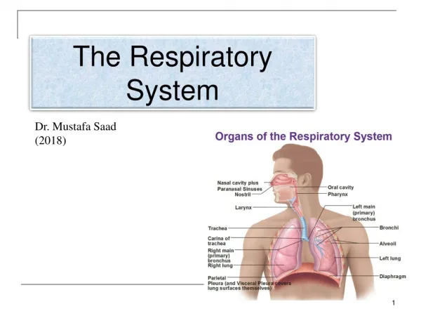
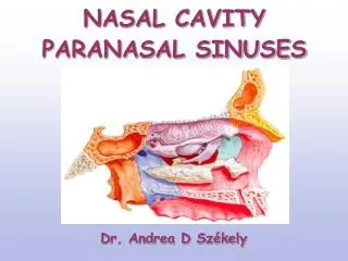
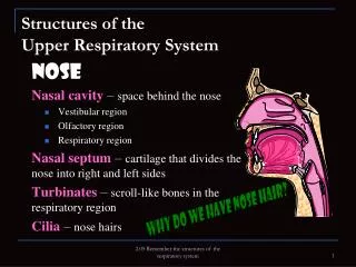
Nose • Nose,is the only visible part of the respiratory system and serves as the entrance to the respiratory tract • The nose has two cavities, separated from one another by a wall called the nasal septum. • The external openings, known as external (anterior) nares ornostrils,lead to the nasal cavities. root tip ala septum external nares • Formed: • aboveby bony skeleton & • belowby plates of hyaline cartilage.
Nasal Cavity • Extends from the external (anterior) nares to the posterior nares (choanae). • Divided into right & left halves by the nasal septum. • Each half has a: • Roof • Lateral wall • Medial wall (septum) • Floor
Roof Narrow & formed (from behind forward) by the: Body of sphenoid. Cribriform plate of ethmoid bone. Frontal bone. Nasal bone & cartilage • Medial Wall (Nasal Septum) • Osteo-cartilaginous partition. • Formed by: • Perpendicular plate of ethmoid bone. • Vomer. • Septal cartilage. 3 2 4 1 • Floor • Formed by the hard (bony) palate. • Separates it from the oral cavity.
Lateral Wall • Shows three horizontal bony projections, the superior,middle&inferiorconchae. • The cavity below each concha is called a meatus and are named as superior, middle & inferior corresponding to the conchae. • The small space above the superior concha is the sphenoethmoidal recess. The conchae are covered by respiratory epithelium and thus increase the surface area of the nasal cavity.
The recess & meati receive the openings of the: Paranasal sinuses. Nasolacrimal duct.
Nerve Supply Olfactory nerves. Ophthalmic& Maxillary divisions. Autonomic fibers. • Arterial Supply: • Branches of the • Maxillary, • Facial & • Ophthalmic arteries. Venous Drainage: • Facial, • Ophthalmic • Spheno-palatine veins. LymphaticDrainage:The lymphatics from the: • The submandibular lymph nodes. • The upper deep cervical lymph nodes.
Paranasal Sinuses • Air filled cavities located in the bones around the nasal cavity: ethmoid, sphenoid, frontal bones & maxillae. • Lined by respiratory mucosa which is continuous with the mucosa of the nasal cavity. • Drain into the nasal cavity. Functions • Lighten the skull. • Act as resonant chambers for speech. • Air conditioning: The respiratory mucosal lining helps in warming, cleaning and moistening the incoming air.
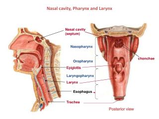
Pharynx • Muscular tube lying behind the nasal cavity, oral cavity & larynx. • Extends from the base of the skull to level of the 6th cervical vertebra, where it is continuous with the esophagus • Divided into three parts: • Nasopharynx: • Superior part, communicates with the nasal cavity through posterior nasal apertures. • Oropharynx: • Middle part, communicates with the oral cavity through theoro-pharyngeal isthmus. • Laryngopharynx: • Inferior part, communicates with the larynx through the laryngeal inlet. Nasal cavity Naso- pharynx * Oro- pharynx Oral cavity * Laryngo- pharynx larynx Esophagus
Nasopharynx • Extends from the base of skull to the soft palate. • Contains Pharyngeal tonsils (adenoides) in its roof. • Lateral wall shows: • Opening of auditory tube. • Tubal elevation (produced by posterior margin of the auditory tube). • Tubal tonsil. • Salpingopharyngeal fold (raised by salpingo-pharyngeus muscle).
Oropharynx • Extends from soft palate toupper border of epiglottis. • Lateral wall shows: • Palatopharyngeal folds. • Palatpglossal fold • Palatine tonsil located between them in a depression called the ‘tonsillarfossa’. Palatoglossal fold Palato-pharyngeal fold Tonsillar fossa
Laryngopharynx • Extends from upper border of epiglottis to lower border of cricoid cartilage. • A small depression situated on either side of the laryngeal inlet is called ‘piriformfossa’. • It is a common site for the lodging of foreign bodies. • Branches of internal laryngeal (& recurrent laryngeal) nerve lie deep to the mucous membrane of the fossa and are vulnerable to injury during removal of a foreign body.
Muscles of Pharynx • The muscles of the pharynx are arranged in circular and longitudinal layers • Circular (Constrictor) • Three muscles, overlap each other: Superior, Middle & Inferior • Propel the bolus of food down into the esophagus • Longitudinal Muscles • Three muscles: • Stylopharyngeus • Salpingopharyngeus • Palatpharyngeous • Elevate the larynx & pharynx during swallowing S M I
Nerve Supply • Sensory: • Nasopharynx: Maxillary nerve • Oropharynx: Glossopharyngeal nerve • Laryngopharynx: Vagus nerve • Motor Nerve Supply: • All the muscles of pharynx, except the stylopharyngeus, supplied by the pharyngeal plexus. • Stylopharyngeus is supplied by the glossopharyngeal nerve Arterial supply: From branches of: • Ascending pharyngeal artery • Ascending palatine artery • Facial artery • Maxillary artery • Lingual artery • The Veins drain into pharyngeal venous plexus, which drains into the internal jugular vein • The lymphatics drain into • The deep cervical lymph node • Rretropharyngeal , • Paratracheallymph nodes