
Respiratory System
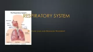
Respiratory System. BY: Chris Lyles and Drakailen Woodruff. Parts Of The Respiratory system. Nose and Nasal Cavity Mouth Pharynx Larynx Trachea Bronchi and Bronchioles Lungs Diaphragm. Nose and Nasal Cavity.

Respiratory System
E N D
Presentation Transcript
Respiratory System BY: Chris Lyles and Drakailen Woodruff
Parts Of The Respiratory system • Nose and Nasal Cavity • Mouth • Pharynx • Larynx • Trachea • Bronchi and Bronchioles • Lungs • Diaphragm
Nose and Nasal Cavity The nose and nasal cavity form the main external opening for the respiratory system and are the first section of the body’s airway—the respiratory tract through which air moves. The nose is a structure of the face made of cartilage, bone, muscle, and skin that supports and protects the anterior portion of the nasal cavity. The nasal cavity is a hollow space within the nose and skull that is lined with hairs and mucus membrane. The function of the nasal cavity is to warm, moisturize, and filter air entering the body before it reaches the lungs.
Mouth The mouth, also known as the oral cavity, is the secondary external opening for the respiratory tract. Most normal breathing takes place through the nasal cavity, but the oral cavity can be used to supplement or replace the nasal cavity’s functions when needed. Because the pathway of air entering the body from the mouth is shorter than the pathway for air entering from the nose, the mouth does not warm and moisturize the air entering the lungs as well as the nose performs this function. The mouth also lacks the hairs and sticky mucus that filter air passing through the nasal cavity. The one advantage of breathing through the mouth is that its shorter distance and larger diameter allows more air to quickly enter the body.
Pharynx The pharynx, also known as the throat, is a muscular funnel that extends from the posterior end of the nasal cavity to the superior end of the esophagus and larynx. The pharynx is divided into 3 regions: the nasopharynx, oropharynx, and laryngopharynx. The nasopharynx is the superior region of the pharynx found in the posterior of the nasal cavity. Inhaled air from the nasal cavity passes into the nasopharynx and descends through the oropharynx, located in the posterior of the oral cavity. Air inhaled through the oral cavity enters the pharynx at the oropharynx. The inhaled air then descends into the laryngopharynx, where it is diverted into the opening of the larynx by the epiglottis.
Larynx The larynx, also known as the voice box, is a short section of the airway that connects the laryngopharynx and the trachea. The larynx is located in the anterior portion of the neck, just inferior to the hyoid bone and superior to the trachea. Several cartilage structures make up the larynx and give it its structure. The epiglottis is one of the cartilage pieces of the larynx and serves as the cover of the larynx during swallowing. Inferior to the epiglottis is the thyroid cartilage, which is often referred to as the Adam’s apple as it is most commonly enlarged and visible in adult males. The thyroidholds open the anterior end of the larynx and protects the vocal folds. Inferior to the thyroid cartilage is the ring-shaped cricoid cartilage which holds the larynx open and supports its posterior end. In addition to cartilage, the larynx contains special structures known as vocal folds, which allow the body to produce the sounds of speech and singing.
Trachea The trachea, or windpipe, is a 5-inch long tube made of C-shaped hyaline cartilage rings lined with pseudostratified ciliated columnar epithelium. The trachea connects the larynx to the bronchi and allows air to pass through the neck and into the thorax. The rings of cartilage making up the trachea allow it to remain open to air at all times. The open end of the cartilage rings faces posteriorly toward the esophagus, allowing the esophagus to expand into the space occupied by the trachea to accommodate masses of food moving through the esophagus.
Bronchi and Bronchioles At the inferior end of the trachea, the airway splits into left and right branches known as the primary bronchi. The left and right bronchi run into each lung before branching off into smaller secondary bronchi. The secondary bronchi carry air into the lobes of the lungs—2 in the left lung and 3 in the right lung. The secondary bronchi in turn split into many smaller tertiary bronchi within each lobe. The tertiary bronchi split into many smaller bronchioles that spread throughout the lungs. Each bronchiole further splits into many smaller branches less than a millimeter in diameter called terminal bronchioles. Finally, the millions of tiny terminal bronchioles conduct air to the alveoli of the lungs.
Lungs Thelungs are a pair of large, spongy organs found in the thorax lateral to the heartand superior to the diaphragm. Each lung is surrounded by a pleural membrane that provides the lung with space to expand as well as a negative pressure space relative to the body’s exterior. The negative pressure allows the lungs to passively fill with air as they relax. The left and right lungs are slightly different in size and shape due to the heart pointing to the left side of the body. The left lung is therefore slightly smaller than the right lung and is made up of 2 lobes while the right lung has 3 lobes. The interior of the lungs is made up of spongy tissues containing many capillaries and around 30 million tiny sacs known as alveoli. The alveoli are cup-shaped structures found at the end of the terminal bronchioles and surrounded by capillaries. The alveoli are lined with thin simple squamous epithelium that allows air entering the alveoli to exchange its gases with the blood passing through the capillaries.
Muscles of Respiration Surrounding the lungs are sets of muscles that are able to cause air to be inhaled or exhaled from the lungs. The principal muscle of respiration in the human body is the diaphragm, a thin sheet of skeletal muscle that forms the floor of the thorax. When the diaphragm contracts, it moves inferiorly a few inches into the abdominal cavity, expanding the space within the thoracic cavity and pulling air into the lungs. Relaxation of the diaphragm allows air to flow back out the lungs during exhalation.
Work Cited http://www.innerbody.com/anatomy/respiratory Images: https://www.google.com/search?q=respiratory+system+detailed&newwindow=1&site=webhp&source=lnms&tbm=isch&sa=X&ei=rVE5U_XRFrK-sQTzx4HYBA&ved=0CAgQ_AUoAQ&biw=1920&bih=979#imgdii=_


