Download
1 / 25
270 likes | 1.6k Vues
The Respiratory System. Dr. Mustafa Saad (2018). Divisions of the Respiratory System. Structurally: (according to embryological origin) Upper respiratory system Nose, pharynx and associated structures Lower respiratory system Larynx, trachea, bronchi and lungs Functionally:

E N D
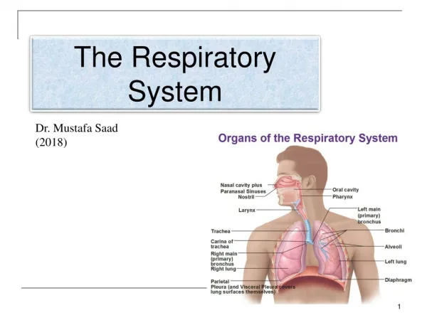
The Respiratory System Dr. Mustafa Saad (2018)
Divisions of the Respiratory System • Structurally: (according to embryological origin) • Upper respiratory system • Nose, pharynx and associated structures • Lower respiratory system • Larynx, trachea, bronchi and lungs • Functionally: • Conducting zone – conducts air to lungs • Nose, pharynx, larynx, trachea, bronchi, bronchioles and terminal bronchioles • Respiratory zone – site of gas exchange • Respiratory bronchioles, alveolar ducts, alveolar sacs, and alveoli
The Nose • Consists of the External nose and Internal nose. • External nose – portion visible on face • Formed of a small bony part – the nasal bones (bridge of the nose) and a larger cartilaginous part. All covered by muscles and skin. • Its openings are called the external nares or nostrils. • The area just inside the nostrils is called the vestibule. Its lower half is lined by skin with hairs, sweat and sebaceous glands. Fig.2: The external nose.
Internal nose – large cavity beyond the vestibule • Extends from vestibule to the internal nares or choanae. • Nasal cavity divided by nasal septum. • The lateral wall has projections that increase the surface area called conchae. Between these conchae and the lateral wall are small passages called meatuses. • Ducts from paranasal sinuses and nasolacrimal ducts open into the meatuses of the internal nose. • Olfactory epithelium responsible for the sense of smell lines the roof of the nasal cavity.
Location of the Olfactory epithelium Fig.3: The internal nose (pink boxes).
Functions of the nose: Warms air coming through the nose due to the rich capillaries of the mucosa of the nose. Moistens incoming air by the mucous secreted by goblet cells of the mucosa. Traps and removes dust particles by cilia and hair. Affects quality of voice. Olfactory epithelium responsible for the sense of smell.
The Pharynx • Funnel shaped tube that starts at internal nares and extends to the level of the cricoid cartilage of larynx (C6 vertebra). • Contraction of skeletal muscles assists in swallowing. • Functions • Passageway for air and food • Resonating chamber • Houses tonsils • 3 anatomical regions • Nasopharynx • Oropharynx • Laryngopharynx Fig.4: Regions of the pharynx.
The Nasopharynx Fig.5: The auditory tube. • Located posterior to the nasal cavities and above the soft palate and inferiorly opens into the oropharynx. It’s a passageway for air. • Its lateral walls exhibit the opening of the auditory (Eustachian) tubes. This tube connects the middle ear with the pharynx balancing air pressure around the eardrum. • The posterior wall of the nasopharynx has a collection of lymphoid tissue called the pharyngeal Tonsil (adenoid).
The Oropharynx • Located posterior to the oral cavity with which it communicates by an opening called fauces. It allows passage of both air and food. Inferiorly, it extends to the level of the hyoid bone. • Anteriorly, in the posterior aspect of the tongue, there’s the lingual tonsil. Anterolaterally, we have the two palatine tonsils. The Laryngopharynx • Located posterior to the larynx with which it communicates. It begins at the level of the hyoid bone and ends at the level of the cricoid cartilage (C6) where it becomes continuous with esophagus. • Muscles of the pharynx play a part in the process of deglutition. These muscles are supplied by the phryngeal plexus derived from the glossopharyngeal (IX) and vagus (X) nerves
Fig.7: The oropharynx (blue boxes) and the laryngopharynx (brown box).
The Larynx • Short passageway connecting laryngopharynx with trachea that’s responsible for the production of sound. Its inlet also provides a protective cover for the airway passages. • It lies in the midline of the neck opposite C4-C6 vertebrae. At the level of C6, it becomes continuous with the trachea. • Important relations: • Anteriorly, it’s covered by the infrahyoid muscles. • Posteriorly, laryngopharynx. • Laterally, the thyroid gland and the great blood vessels of the neck. • Superiorly, the hyoid bone. • Inferiorly, the trachea. Fig.8: Important relations of the larynx.
The framework of the larynx is formed of cartilages that are held together by ligaments and membranes, moved by muscles and lined by mucous membrane. Anterior view Posterior view Fig.9: Structure of the larynx.
Cartilages of the Larynx Thyroid: single V-shaped cartilage. The largest and forms prominence of the neck called Adam’s Apple. Cricoid: a single ring shaped cartilage. The lower boundary of the larynx. Epiglottis: a leaf-shaped single cartilage that’s present at the inlet of the larynx. Closes the larynx when the person swallows. Arytenoid: a pair of pyramidal cartilages to which the vocal cords are attached. Movement of these cartilages leads to movement of the cords. Corniculate cartilages (pair). Cuneiform cartilages (pair). Fig.10: Cartilages of the larynx.
Structures of Sound Production • Mucous membrane of larynx forms 2 folds/ligaments: • Vestibular folds(false vocal cords) – superior pair • Function in holding breath against pressure in thoracic cavity as in heavy weight lifting. • Vocal folds (true vocal cords) – inferior pair • Contain elastic tissue. • Muscle contraction pulls elastic ligaments which stretch vocal folds. • Passing air vibrates the cords and produce sound. • Folds can move apart or together, elongate or shorten. • Muscles of the larynx are Intrinsic or Extrinsic. The intrinsic muscles are supplied by branches of the vagus (X) nerve.
Fig.11: Different positions of the vocal cords. Below, sagittal section of the larynx showing the two folds.
The Trachea • A Tubular passage of air that extends from the larynx to superior border of T5 where it divides into right and left primary bronchi. • Wall is formed of 4 layers: • Mucosa • Submucosa • Hyaline cartilage • Adventitia (Connective tissue) • 16-20 C-shaped rings of hyaline cartilage: • Open part faces esophagus. • The opening of the C is bridged by a fibromuscular membrane formed of elastic fibers and the smooth Trachealismuscle. Fig.12: Wall of the trachea.
Important relations of the trachea • Anterior: • Infrahyoid muscles • Thyroid gland • Lateral: • Thyroid gland • Internal jugular vein • Common carotid artery • Vagus nerve • Posterior: • Recurrent laryngeal nerve • Esophagus and • Vertebral column Fig.13: Relations of the trachea.
The Bronchial Tree • Right and left primary bronchi pass to the corresponding lung. • Carina – angle between the two main bronchi. • Divide to form bronchial tree: • Secondary lobar bronchi (one for each lobe), tertiary (segmental) bronchi, bronchioles, terminal bronchioles • Structural changes with branching: • Mucous membrane changes. • Cartilage decreases. As it decreases, smooth muscle increases. • Terminal bronchioles will form Respiratory bronchioles. At this point gas exchange begins to occur.
The Lungs • Two cone shaped organs separated from each other by the heart and other structures in the mediastinum. • Each lung is enclosed by the double-layered pleura (serous membrane): • Parietal pleura – lines wall of thoracic cavity. • Visceral pleura – covers the lungs themselves. • Pleural cavity is space between layers • Pleural fluid reduces friction • Lungs receive blood from: • Pulmonary artery - deoxygenated blood. • Bronchial arteries – oxygenated blood to supply nutrients for cells.
Alveoli • Cup-shaped outpouching • 2 types of alveolar epithelial cells: • Type I alveolar cells – main site of gas exchange. • Type II alveolar cells – secrete surfactant factor which reduces tendency to collapse. • Respiratory membrane: a very thin cellular membrane separating air from blood and across which gas exchange occurs: • Alveolar wall – type I alveolar cells • Epithelial basement membrane • Capillary basement membrane • Capillary endothelium


