Download
1 / 63
670 likes | 1.11k Vues
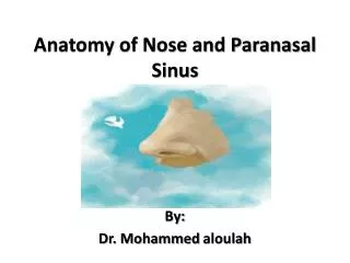
Nose and paranasal sinuses. Ehab ZAYYAN, MD, PhD. External nose. Root and tip Nostrils (nares): are the external orifices of the nose. Bounded laterally by the ala and medially by the nasal septum Nasal skeleton: Nasalis bones Frontal processes of the maxillae

E N D
Nose and paranasal sinuses Ehab ZAYYAN, MD, PhD
External nose • Root and tip • Nostrils (nares): are the external orifices of the nose. Bounded laterally by the ala and medially by the nasal septum Nasal skeleton: • Nasalis bones • Frontal processes of the maxillae • Nasal part of the frontal bone • Upper and lower nasal cartilages • Septal cartilage
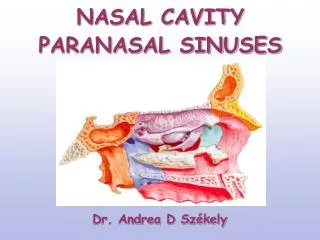
Nasal cavity • From the nostrils till the choana • Divided into right and left by the nasal septum • Floor: palatine process of the maxilla and horisontal plates of the palatine bones • Roof: body of the sphenoid, cribriform plate of the ethmoid, frontal bone, nasal bone, nasal cartilages
Lateral nasal wall • Superior, middle and inferior conchae. The area below each concha is called meatus
The middle meatus is continuous in front with a depression called the atrium. • Within the nostril in front of the atrium is the vestibule. It is lined by modified skin and short curved hair called the vibrissae
The mucous membrane lines the nasal cavity except the vestibule. There are two types of mucous membranes: 1. Olfactory mucosa 2. Respiratory mucosa
1. olfactory mucous membrane: lines the superior surface of the superior concha and the sphenoethmoidal recess. It also lines the corresponding part of the nasal septum
The axons of the fibers pass through the openings in the cribriform plates of the ethmoid bone and end in the olfactory bulb
2. The respiratory mucous membrane: • Lines the rest of the nasal cavity. • Its function is to warm, moisten and clean the inspired air. • Warming: venous plaxus in the submucosa • Moisture: mucous by the glands and goblet cells • Ciliary action….
Nerve supply of the nasal cavity • The olfactory nerve: arise from special olfactory cells in the olfactory mucous membrane • Nerves of general sensation: derived from opthalmic and maxillary divisions of the trigeminal nerve: • - anterior ethmoid nerve • - nasal, nasopalatine , greater and lesser palatine nerves of the pterygopalatine ganglion
Sympathetic innervation: • T1-T3 → SCSG → ICA plexus → deep petrosal nerve → vidian nerve → PPG Parasympathetic innervation: • Superior salivatory nucleus → facial nerve → GSPN → vidian nerve → PPG → nasal branches through the sphenopalatine foramen
Blood supply of the nasal cavity ECA 1. Sphenopalatine artery (maxillary artery) 2. Greater palatinal artery branch in the incisive canal (maxillary artery) 3. Superior labial branch of the facial artery ICA 4. Anterior ethmoid artery (ophthalmic artery) 5. Posterior ethmoid artery (ophthalmic artery)
Kiesselbach area (Little area) of the nasal septum • The area of the septum about 1 cm posterior to the nares. It is very rich in blood supply and a common site of epistaxis. • Anterior ethmoid artery • Sphenopalatine artery • Greater palatinal artery • Septal branch of facial artery
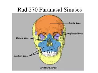
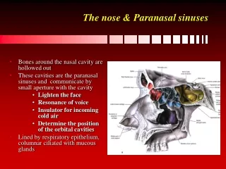
Paranasal sinuses • Cavities found in the interior of the maxilla, frontal,sphenoid and ethmoid bones. • Lined with mucoperiostium and filled with air. • The mucous produced by the glands in the mucous membranes is moved into the nose by the ciliary action of the columnar cells. • The function of the sinuses is to act as resonators of the voice and to reduce the weight of the skull.