Download
1 / 67
2.79k likes | 7.39k Vues
Anatomy and relevant anatomical variants in nasal and paranasal sinuses CT scan. CERTIFICATE OF MERIT RSNA 2003. P. Loubeyre 1 MD & J.S Lacroix 2 MD, PhD 1 Radiology Department, 2 Rhinology-Olfactology Unit Hôpitaux Universitaires de Genève CH- 1211 Genève 14 Switzerland.

E N D
Anatomy and relevant anatomical variants in nasal and paranasal sinuses CT scan CERTIFICATE OF MERIT RSNA 2003 P. Loubeyre1 MD & J.S Lacroix2 MD, PhD 1 Radiology Department, 2 Rhinology-Olfactology Unit Hôpitaux Universitaires de Genève CH- 1211 Genève 14 Switzerland
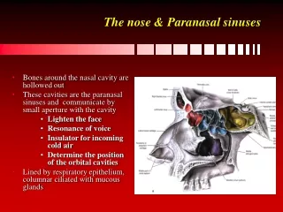
Anatomic variants Septal deviation (dia 40) Septal spurs (dia 41) Middle turbinate (dia 42-44) Superior turbinate (dia 45) Uncinate process (dia 46) Frontal bulla cell (dia 47) Frontal sinus extension (dia 48) Agger nasi cell (dia 49) Maxillary sinus recesses (dia 50) Sphenoid sinus recesses (dia 51-53) Intersinus sphenoid septation (dia 54) Surgical risks Vulnerability of the carotid canal (dia 55-57) Vulnerability of the optic nerve (dia 58-61) Vulnerability of the orbit (dia 62-65) Vulnerability of the nasolacrymal duct (dia 66) Vulnerability of anterior skull base (dia 67) Sinusitis – Imaging technique (3-10) Anatomy Paranasal sinuses (dia 11-12) Ostiomeatal unit (dia 13-16) Uncinate process (dia 17-19) Ethmoid bulla (dia 20-22) Middle turbinate (dia 23,24) Ethmoid infundibulum (dia 25) Frontal recess (dia 26-28) Sphenoethmoidal recess (dia 29,30) Posterior nasal fontanel (dia 31) Posterior choana (dia 32) Nasal septum (dia 33) Nasolacrymal duct (dia 34) Anterior skull base (dia 35-38) Some articles to read Mafee MF. Endoscopic sinus surgery: role of the radiologist. AJNR1991;157:855-60 Earwaker J. Anatomic variants in sinonasal CT. Radiographics 1993;13:381-415 Rao VM, El-Noueam KI. Sinonasal imaging. Radiologic Clinics of North America 1998;36(5):921-39 Zeifer B. Update on sinonasal imaging. Neuroimaging Clinics of North America 1998;8(3):607-30
Mucociliary clearance impairment Mucous accumulation increases the risk of infection or chronic inflammation
Mucociliary transport : anatomy of the drainage pathway of the paranasal sinuses Ostio-meatal complex
Obstruction Normal mucociliary clearance or Normal mucosal thickening during nasal cycle Contact of two opposing mucosal surfaces (ex: anatomic variants) Mucous accumulation increases risk of infection
Chronic rhinosinusitis unresponsive to medical management The aim of nasal and paranasal sinuses imaging is to provide a surgical road map delineating the anatomy, defining the obstructive lesions, and noting the anatomical factors that may predispose impaired mucociliary clearance and per operative complications CT High spatial resolution Minute bony details and adjacent soft tissue structures Pre-operative investigation for patients undergoing endoscopic sinus surgery Rhinosinusitis is a medical diagnosis, not a radiologic diagnosis
Supine position Confortable position console Coronal, sagittal or oblique reformations Real time image-guided anatomic localization during endoscopic surgery 1-mm-thick overlapping axial slices
Medical treatment should be performed before CT scan To reduce transient acute inflammatory or infectious mucosal changes Nasal lavages Topical corticosteroids Antibiotics Mucolytic agents CT scan should be scheduled after completion of medical treatment
Contrast-enhanced CT ? .Anatomy is adequately assessed without the use of IV injected iodinated contrast material .Iodine injection does not allow for discriminating an inflammatory disease from a tumoral process no Initial images or clinical symptoms suggest intracranial complications ofa chronicinflammatory sinus disease yes MRI
CT imaging 1. Obstruction of the drainage pathways or anatomic variants that may compromise already narrow drainage pathways 2. Identification of critical anatomic areas where anatomic variants pose special risks during sinus surgery 3. Local extension of disease 4. Complications 1+2 : Preoperative CT scan as a road map for endoscopic sinus surgery
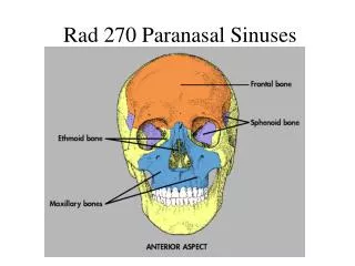
Coronal CT Maxillary sinuses Ethmoid sinus Nasal cavity
Sagittal CT Frontal sinuses Ethmoid sinus Sphenoid sinus Nasal cavity
Ostio-meatal unit Coronal CT Sagittal CT Middle nasal meatus (air channel medial to uncinate process and lateral to the middle turbinate)
Ostio-méatal unit Drainage from frontal, maxillary and anterior ethmoid sinuses Obstruction Frontal sinusitis Maxillary sinusitis Anterior ethmoid sinusitis Sagittal view
Ostio-meatal unit Coronal CT Ethmoid bulla (EB) Orbit EB Middle turbinate Middle meatus Uncinate process Axial CT
Ostio-meatal unit Maxillary ostium Ethmoid infundibulum (posterior) Hiatus semilunaris Middle meatus Coronal CT
Uncinate process Thin-curved bony lamina of variable height from the lateral side of the ethmoid labyrinth, that forms a portion of the lateral nasal wall. Coronal CT EB Lamina papyracea Supero-anterior attachment to lamina papyracea in 50% Inferior attachment to the neck of the inferior turbinate
Uncinate process Coronal CT Sagittal CT Postero-lateral attachment to the roof of the maxillary sinus
Uncinate process Coronal CT Sagittal CT EB Anterior-most ethmoid cell: agger nasi cell Lacrymal bone Anterior attachment of the uncinate process
Ethmoid bulla (EB) Most posterior of all anterior ethmoid air cells, roof of the hiatus semilunaris and posterior ethmoid infundibulum Coronal CT Sagittal CT EB Anterior ethmoid drainage Middle meatus Anterior ethmoid cells can drain into the middle meatus via the ethmoid bulla
Ethmoid bulla Orbit EB Uncinate process Normal-sized bullae Large bullae Hypoplasic bullae Acording to John Earwaker. Anatomic variants in sinonasal CT.Radiographics 1993;13:381-415
Coronal CT Different ethmoid bullae
Middle turbinate Coronal CT Coronal CT Posterior ethmoid cell EB Bulb It attaches posteriorly and laterally to the lamina papyracea: Basal (ground) lamella It attaches superiorly to the cribriform plate: Medial lamella
Coronal CT Middle turbinate Sagittal CT Posterior ethmoid air cells Anterior ethmoid air cells Posterior ethmoid Anterior ethmoid EB Basal lamella
Ethmoid infundibulum Anterior ethmoid cells drainage Frontal sinus drainage (25%) Anterior ethmoid cells can directly drain into middle meatus Coronal CT
Frontal recess Communication between frontal sinus and nasal cavity. It is not strictly a duct but a channel located between anterior ethmoid cells. Variety of configurations. Sagittal CT Coronal CT Frontal sinus Frontal ostium Agger nasi cell Anterior middle meatus
Coronal CT Frontal sinus drainage pathways according to supero-anterior attachment of the uncinate plate 50% Attachment to the lamina papyracea Frontal drainagein the medial meatus 25% Attachment to the skull base Frontal drainageinto the ethmoid infundibulum 25% Attachment to the neck of the middle turbinate Frontal drainage into the ethmoid infundibulum or into an anterior ethmoid cell
Frontal sinus outflow obstruction Frontal sinusitis Sagittal view
Sphenoethmoidal recess Posterior ethmoid and sphenoid sinus drainages Axial CT Sagittal CT Sphenoid sinus Sphenoid sinus ostium Superior nasal meatus
Sphenoethmoidal recess obstruction Posterior ethmoid sinusitis Sphenoid sinus sinusitis
Posterior nasal fontanel Coronal CT coronal Axial CT Area of the lateral wall of the nose immediately behind the posterior attachment of the uncinate plate. Consists of mucous membrane only, without bony support. Accessory maxillary ostium is frequently found through the posterior nasal fontanel (15-40%)
Posterior choana Coronal CT Sagittal CT Nasopharynx
Nasal septum Coronal CT Sagittal view Perpendicular plate of the ethmoid post ant Septal cartilage vomer Chondrovomeral junction
Nasolacrymal duct 2 Axial CT 1 Coronal CT 3 4 Inferior meatus
Anterior skull base Coronal CT Axial CT Crista galli Cribriform plates (floor of the olfactive fossa) Ethmoid roof (fovea ethmoidalis)
Ethmoid roof Axial CT Sagittal CT
Lateral lamella (point of structural weakness in the anterior skull base) Point of structural weakness during turbinectomy Coronal CT Ethmoid roof Medial lamella Cribriform plate Middle turbinate 1-16 mm length Dehiscent in 15% of specimens Anterior ethmoidal artery
Anterior ethmoidal artery Coronal CT Coronal CT Olfactory fossa Lateral lamella Medial lamella of Middle turbinate The anterior ethmoidal artery - branch of the ophtalmic artery - exits the orbit through the anterior ethmoidal foramen and enters the olfactory fossa at the point of attachment of the middle turbinate to the cribriform plate
Anatomic variants Very frequently noted The presence of anatomic variants, singly or in combination, does not represent a disease state per se Equal prevalence of patients with and without sinus disease in the presence of the same anatomic variant
Septal deviation Coronal CT Coronal CT Hypoplasic middle turbinate Large middle turbinate Middle meatus Inferior meatus
Septal spurs Axial CT Coronal CT Coronal CT Bridging spur Frequently encountered at the junction of the perpendicular plate of the ethmoid and the vomer May impige on and invaginate the middle or inferior turbinates. When the turbinate mucosa swells with the normal nasal cycle or inflammation, it is impaled on the spur, setting up a cycle of facial pain or headache. A septal spur occasionally produces a complete bridge.
Size variations of middle turbinate Coronal CT Coronal CT Ostio-méatal complex compromise? Turbinate septal contact when the turbinate mucosa swells with the normal nasal cycle or inflammation
Pneumatized middle turbinate Axial CT Coronal CT Air cell in the vertical lamella: concha neck air cell Air cell in the bulbous segment: concha bullosa cell
Paradoxical middle turbinate Coronal CT Usually Convex configuration medially Concave configuration laterally Paradoxical turbinate Concave configuration medially Convexe configuration laterally
Pneumatized superior turbinate Coronal CT A cause of migraine headache? Enlargement of the superior turbinate due to pneumatization, with accompanying mucosal contact, acts as a mechanical stimulus initiating an axon reflex with resultant referred pain?
Uncinate process Height : 1-4 mm Length : 14-22 mm Coronal CT Coronal CT Medially rotated uncinate Pneumatized
Frontal bulla cell (suprabullar cell) Anterior ethmoid air cell extending upwards (intramural ethmoid air cell) sagittal coronal axial Frontal recess May be small and impiges only on the floor of the frontal sinus. May elevate and narrows the frontal recess May be prominent and bulges into the frontal sinus
Frontal sinus extension Pneumatization of the orbital plate of the frontal bone ant post Coronal CT Frontal sinus No frontal sinus drainage compromise
Agger nasi (AN) cell Coronal CT Sagittal CT AN Lacrymal fossa Frontal recess The most anterior of the ethmoid cells. Forms the floor of the frontal recess. It reaches the lacrimal fossa inferiolaterally, and is anterolaterally arched by the nasal bone. A large agger nasi can impige on and distort the frontal recess. Its posterior-medial wall usually gives rise to the anterior uncinate process
Maxillary sinus recesses Coronal CT Palatine recess Axial CT Infraorbital recess of The maxillary sinus Coronal CT Axial CT Sagittal CT Alveolar recesses (roots of the premolar and molar teeth) Zygomatic recess