The Nasal Cavity
The Nasal Cavity. Prof. Dr.Mohammed Hisham Al-Muhtaseb. The Nasal Cavity. The first part of the respiratory tract Functions of the respiratory system: 1. Provides for gas exchange 2. Regulates blood ph 3. Filters the inspired air

The Nasal Cavity
E N D
Presentation Transcript
The Nasal Cavity Prof. Dr.Mohammed Hisham Al-Muhtaseb
The Nasal Cavity • The first part of the respiratory tract • Functions of the respiratory system: • 1. Provides for gas exchange • 2. Regulates blood ph • 3. Filters the inspired air • 4. Contains receptors for smell, and produce vocal sounds (phonation) • 5. Excretes small amounts of water and heat
Nose • Devided into : • External nose • Nasal Cavity
External nose • Cartilaginous framework: • 1. Septal cartilage • 2. Lateral nasal cartilage • 3. Alar cartilage • All are plates of hyaline cartilage
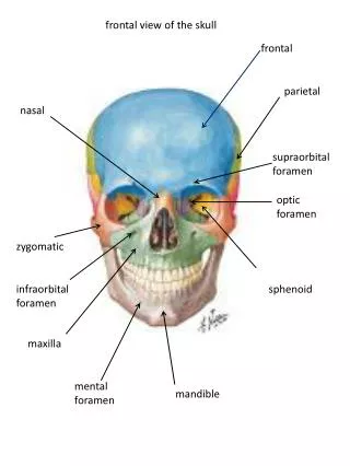
External nose • Bony framework • 1. The nasal bones • 2. Frontal processes of the maxillae • 3. Nasal part of the frontal bone
External Nose • Blood Supply • Branches of the ophthalmic and the maxillary arteries • ala and the lower part of the septum by branches from the facial artery. • Nerve Supply • Infratrochlear and external nasal branches of the ophthalmic nerve • infraorbital branch of the maxillary nerve
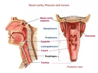
Nasal Cavity • Extends from the nostrils in front to the posterior nasal apertures (choana) • Opens into the nasopharynx • Vestibule is the area of the nasal cavity lying just inside the nostril • Divided into right and left halves by the nasal septum • Septum is made up of the septal cartilage, the vertical plate of the ethmoid, and the vomer.
Nasal Cavity • Functions : • 1. Respiratory • 2. Olfactory • 3. Resonance of voice • 4. drains lacrimal fluid • 5. Protective : • Sneezing • Filtration • Proteolytic enzymes • Warming and moistening the air
Nares • The anterior openings of the nasal cavities • Held open by the surrounding alar cartilages and septal cartilage • Nares are continuously open • Can be widened further by the action of the related muscles of facial expression
Choanae • Openings between the nasal cavities and the nasopharynx • Rigid openings completely surrounded by bone
Nasal Cavity • Boundaries of the cavity : • Floor • Roof • lateral wall • Medial or septal wall.
Floor • The upper surface of the hard palate • which consist of: • 1. Palatine process of the maxilla • 2. Horizontal plate of the palatine bone
Roof • 1. Sloping anterior part: • Nasal spine of the frontal bone and the nasal bones • 2.Horizintal middle part: • The cribriform plate of the ethmoid bone
Roof • 3. Sloping posterior part: • Anterior surface of the sphenoid bone (body) • Ala of the vomer • Vaginal process of the palatine bone
Medial wall • Septal nasal cartilage anteriorly • Posteriorlyvomer and the perpendicular plate of ethmoid bone
Lateral Wall • Complex and formed by bone, cartilage, and soft tissues • Bony support : • Ethmoidal labyrinth and uncinate process • Perpendicular plate of the palatine bone • Medial plate of the pterygoid process • Medial surfaces of the lacrimal bones and maxillae • Inferior concha
Lateral Wall • Parts: • 1. Vestibule is the area of the nasal cavity lying just inside the nostril • Covered with skin and contains thick hairs (vibrissae) • 2. Antrum (atrium) • 3. Posterior part contain 3 conchae, 3 meatuses, and one recess.
Mucosa • lined with respiratory mucous membrane • Except : • 1. The vestibule is lined with modified skin and has coarse hairs • 2. Above the superior concha is lined with olfactory mucous membrane and contains nerve endings
Function of Mucous Membrane • large plexus of veins in the submucous connective tissue is present in the respiratory region. • Warm blood in the venous plexuses serves to heat up the inspired air as it enters the respiratory system • Mucous traps foreign particles and organisms in the inspired air
Choncae • All Choncae extend medially across the nasal cavity, separating it into four air channels: • Inferior, Middle, and Superior meatus, and a Spheno-ethmoidal recess • Anterior end of each concha curves inferiorly to form a lip that overlies the end of the related meatus
Choncae • lateral wall of the middle meatus elevates to form the dome-shaped Ethmoidal bulla • Formed by the underlying middle ethmoidal cells, which expand the medial wall of the ethmoidal labyrinth. • Inferior to the ethmoidal bulla is a curved gutter (the Hiatus semilunaris), • Formed by the mucosa covering the lateral wall • Defect in the bony wall between the ethmoidal bulla above and the uncinate process below.
Choncae • Anterior end of the hiatus semilunaris forms a channel (the Ethmoidal infundibulum), • Curves upwards and continues as the Frontonasal duct through the anterior part of the ethmoidal labyrinth to open into the frontal sinus.
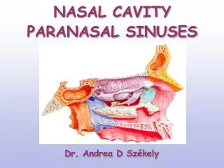
Paranasal Sinuses and Their Site of Drainage Into the Nose • The nasolacrimal duct and most of the paranasal sinuses open onto the lateral wall of the nasal cavity • 1. Maxillary sinus • Middle meatus through hiatus semilunaris • 2.Frontal sinuses • Middle meatus via infundibulum and frontonasal duct • 3. Sphenoidal sinuses • Sphenoethmoidal recess
Paranasal Sinuses and Their Site of Drainage Into the Nose • Ethmoidal sinuses • 1. Anterior group • Infundibulum and into middle meatus • 2. Middle group • Middle meatus on or above bulla ethmoidalis • 3.Posterior group • Superior meatus • Nasolacrimal duct opens onto the lateral wall of the inferior nasal meatus
Blood Supply • Sphenopalatine artery • largest vessel supplying the nasal cavity • Terminal branch of the maxillary artery in the pterygopalatine fossa • Enters the nasal cavity by passing medially through the sphenopalatine foramen
Branches • 1. Posterior lateral nasal branches • Short sphenopalatine artery • supply a large part of the lateral wall (post.superior quadrant)
Branches • 2.. Posterior septal branches • Long sphenopalatine • pass over the roof of the cavity and onto the nasal septum • contribute to the blood supply of the medial wall
Greater palatine artery • Greater palatine artery • Arises in the pterygopalatine fossa as a branch of the maxillary artery • Enters the nasal cavity by passing up through the incisive canal • Supplies the anterior regions of the medial wall and adjacent floor (posterio and anterio-inferior quadrant)
Anterior and posterior ethmoidal arteries • Originate in the orbit from the ophthalmic artery • The anterior ethmoidal artery accompanies the anterior ethmoidal nerve • Descending through a slit-like foramen lateral to the crista galli • Supply the medial (septal) and lateral wall of the nasal cavity (anterior-superior quadrant) • The posterior ethmoidal artery descends into the nasal cavity through the cribriform plate and has branches to the upper parts of the medial and lateral walls.
Superior labial and lateral nasal arteries • Originate from the facial artery on the front of the face • Superior labial gives an alar branch supplies the region around the naris and a septal branch supplies anterior regions of the nasal septum. • lateral nasal arteries supply blood of the external nose • Alar branches pass around the lateral margin of the naris and supply the nasal vestibule.
Epistaxis • Vessels that supply the nasal cavities form extensive anastomoses with each other • in the anterior region of the medial wall there are anastomoses relatively close to the surface(Kiesselbach’s area) • This area is the major site of 'nose bleeds' or epistasxis.
Veins • Veins draining the nasal cavities generally follow the arteries • veins that pass with branches originate from the maxillary artery drain into the pterygoid plexus • veins from anterior regions of the nasal cavities join the facial vein.
Lymphatics • Lymph from anterior regions drains onto the face by passing around the margins of the nares • These lymphatics connect with the submandibular nodes
Innervation • 1. The olfactory nerve [I] for olfaction • 2. Branches of the ophthalmic [V1] and maxillary [V2] nerves for general sensation • 3. Parasympathetic fibers from the facial nerve [VII], Secretomotor innervations of mucous glands
Olfactory nerve [I] • Composed of axons from receptors in the olfactory epithelium at the top of each nasal cavity • Pass superiorly through the cribriform plate to synapse with the olfactory bulb of the brain. • Branches that innervate the nasal cavity: • anterior and posterior ethmoidal nerves, which originate from the nasociliary nerve in the orbit.
Anterior and Posterior ethmoidal nerves • The anterior ethmoidal nerve travels with the anterior ethmoidal artery • It has branches to the medial and lateral wall of the nasal cavity and continues forward on the undersurface of the nasal bone • onto the external surface of the nose by traveling between the nasal bone and lateral nasal cartilage, terminates as the external nasal nerve
Posterior ethmoidal nerve • leaves the orbit through a similar canal in the medial wall of the orbit • Terminates by supplying the mucosa of the ethmoidal cells and sphenoidal sinus • Normally does not extend into the nasal cavity itself.
Branches from the maxillary nerve [V2] • originate in the pterygopalatine fossa just lateral to the lateral wall of the nasal cavity • leave the fossa to enter the nasal cavity by passing medially through the sphenopalatine foramen • 1. Posterior superior lateral nasal nerves pass forward on and supply the lateral wall of the nasal cavity; • 2. Posterior inferior nasal nerves originate from the greater palatine nerve, innervate the lateral wall of the nasal cavity • 3. Anterior superior alveolar branch of the infra-orbital nerve supply the lateral wall near the anterior end of the inferior concha.
Branches from the maxillary nerve [V2] • 4. Largest of these nerves is the nasopalatine nerve, pass through the incisive canal onto the roof of the oral cavity, and terminates by supplying the oral mucosa posterior to the incisor teeth • 5. Posterior superior medial nasal nerves cross the roof to the nasal septum and supply both these regions
Summary for blood supply and innervations • 1. Postero-superior quadrant: • Posterior-superior lateral nerve and vessels (short spheno palatine) • 2. Postero-inferior quadrant: • Greater palatine nerve and vessels • 3. Antero-superior quadrant : • Ant. Ethmoidal nerve (internal and external nerve) and artery • 4. Antero-inferior quadrant : • Ant. Superior alveolar nerve and branches from the facial and greater palatine artery • 5. Nasal septum: • Lower posterior part by the long sphenopalatine nerve • Upper anterior part by the septal branch of the anterior ethmoidal nerve. • Blood supply by the long sphenopalatine artery.
Paranasl sinuses • There are four paranasal air sinuses-the ethmoidal cells, and the sphenoidal, maxillary, and frontal sinuses • All are: • lined by respiratory mucosa, which is ciliated and mucus secreting; • open into the nasal cavities; • innervated by branches of the trigeminal nerve [V].
Paranasl sinuses • Functions: • 1. Resnonance of the voice • 2. Decrease the weight of the skull • 3. Protection
Frontal sinuses • One on each side, seperated by a septum • Triangular in shape and is in the part of the frontal bone under the forehead • Drains onto the lateral wall of the middle meatus via the frontonasal duct, which continues as the ethmoidal infundibulum • Innervated by branches of the supra-orbital nerve from the ophthalmic nerve
Ethmoidal cells • Each cluster of cells is separated from the orbit by the thin orbital plate of the ethmoidal labyrinth • Divided into anterior, middle, and posterior ethmoidal cells • The anterior ethmoidal cells open into the ethmoidal infundibulum or the frontonasal duct; • The middle ethmoidal cells open onto the ethmoidal bulla • The posterior ethmoidal cells open onto the lateral wall of the superior nasal meatus. • Iinnervated by the anterior and posterior ethmoidal branches of the nasociliary nerve from the ophthalmic nerve
Maxillary sinuses • The largest of the paranasal sinuses and completely fill the bodies of the maxillae • Pyramidal in shape • Apex directed laterally • Base deep to the lateral wall of the adjacent nasal cavity • Innervated by infra-orbital and alveolar branches of the maxillary nerve • Drains in Middle meatus through hiatus semilunaris (Pad drainage) • Clinical note : Extraction of upper teeth might lead to fistula formation and sinusitis
Maxillary sinuses • Relationships of the maxillary sinus : • Related above to the orbit • Related below to the roots of the upper molar and premolar teeth • Related behind to the infratemporal fossa • Related medially to the lower part of the nasal cavity