Download

1 / 18
250 likes | 2.84k Vues
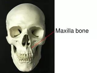
INFRATEMPORAL FOSSA II: MAXILLARY NERVE & VESSELS. Dr. Ahmed Fathalla Ibrahim. THE PTERYGOPALATINE FOSSA. THE PALATE. THE PTERYGOPALATINE FOSSA. BOUNDARIES: ANTERIOR: Maxillary bone POSTERIOR: Pterygoid process of sphenoid bone MEDIAL: Perpendicular plate of palatine bone.

E N D
INFRATEMPORAL FOSSA II: MAXILLARY NERVE & VESSELS Dr. Ahmed Fathalla Ibrahim
THE PTERYGOPALATINE FOSSA • BOUNDARIES: • ANTERIOR: Maxillary bone • POSTERIOR: Pterygoid process of sphenoid bone • MEDIAL: Perpendicular plate of palatine bone
THE PTERYGOPALATINE FOSSA • COMMUNICATIONS: • With infratemporal fossa: through pterygomaxillary fissure • With cranial cavity: through foramen rotundum • With nasal cavity: through sphenopalatine foramen • With orbit: through inferior orbital fissure • With palate: through greater & lesser palatine canals & foramina • With pharynx: through pterygoid canal & palatovaginal canal
THE PTERYGOPALATINE FOSSA • CONTENTS: • Pterygopalatine ganglion • Maxillary nerve • Maxillary artery
MAXILLARY NERVE • ORIGIN: It arises from the trigeminal ganglion in the middle cranial fossa • TYPE OF FIBERS: Pure sensory (general somatic afferent)
MAXILLARY NERVE ZF Zygomatic ZT Meningeal PSA MSA ASA
COURSE OF MAXILLARY NERVE • THE CRANIAL CAVITY: It passes in the lateral wall of cavernous sinus then passes through the foramen rotundum to enter: • THE PTERYGOPALATINE FOSSA: It passes through pterygomaxillary fissure to enter: • THE INFRATEMPORAL FOSSA: It crosses the fossa & passes through the inferior orbital fissure to enter: • THE ORBIT: The nerve is now called theinfraorbital nerve. It runs on the floor of the orbit & passes through the infraorbital groove, canal & foramen to enter: • THE FACE
BRANCHES OF MAXILLARY NERVE • IN THE CRANIAL CAVITY: • Meningeal branch: supplies middle cranial fossa • IN THE PTERYGOPALATINE FOSSA: • Two ganglionic branches connecting the nerve to pterygopalatine ganglion: contain postganglionic parasympathetic fibers to lacrimal gland & sensory fibers from nose, palate & pharynx • IN THE INFRATEMPORAL FOSSA: • Posterior superior alveolar nerve: supplies maxillary sinus & upper molar teeth • Zygomatic nerve: passes through inferior orbital fissure to enter the orbit & divides into zygomaticotemporal & zygomaticofacial nerves
BRANCHES OF INFRAORBITAL NERVE • IN THE INFRAORBITAL CANAL: • Middle superior alveolar nerve: supplies maxillary sinus & upper premolar teeth • Anterior superior alveolar nerve: supplies maxillary sinus & upper canine & incisors • IN THE FACE: • Palpebral: supplies lower eyelid • Nasal: supplies ala of nose • Labial: supplies upper lip
MAXILLARY ARTERY Sphenopalatine ASA PSA Lesser palatine Greater palatine
MAXILLARY ARTERY • ORIGIN:It is the larger of the 2 terminal branches of external carotid artery, behind the neck of mandible (within parotid gland) • COURSE:It is divided into 3 parts: • First part: runs deep to neck of mandible (below auriculotemporal nerve) & emerges from the lower border of lateral pterygoid • Second part: ascends superficial (sometimes deep) to lower head of lateral pterygoid • Third part: passes between the 2 heads of lateral pterygoid then through pterygomaxillary fissure to reach pterygopalatine fossa where it terminates
BRANCHES OF MAXILLARY ARTERY • FROM FIRST PART: • Middle meningeal artery: lies deep to lateral pterygoid, behind the trunk of mandibular nerve, between the 2 roots of auriculotemporal nerve. It passes through foramen spinosum to reach cranial cavity • Inferior alveolar artery: same course & distribution as inferior alveolar nerve • Deep auricular & anterior tympanic arteries: supply external auditory meatus & tympanic membrane
BRANCHES OF MAXILLARY ARTERY • FROM SECOND PART: • Muscular branches: to muscles of mastication • Buccal artery: same course & distribution as buccal nerve
BRANCHES OF MAXILLARY ARTERY • FROM THIRD PART: • Infraorbital artery: same course & distribution as infraorbital nerve • Posterior superior alveolar artery: same course & distribution as posterior superior alveolar nerve • Greater palatine artery: passes through greater palatine canal & foramen to the palate, supplies hard palate & gums, gives lesser palatine artery supplying soft palate & tonsils • Sphenopalatine artery: passes through sphenopalatine foramen to the nose, supplies nasal cavity & nasal septum • Artery of pterygoid canal: supplies auditory tube, tympanic cavity & pharynx • Pharyngeal artery: supplies nasopharynx, sphenoidal air sinuses
PTERYGOID PLEXUS OF VEINS • SITE: It surrounds & lies in substance of lateral pterygoid muscle • TRIBUTARIES: correspond to branches of maxillary artery • TERMINATION: It forms the maxillary vein that passes deep to neck of mandible & unite with superficial temporal vein to form retromandibular vein (inside parotid gland) • COMMUNICATIONS: • With cavernous sinus: by emissary veins through foramen ovale & lacerum • With facial vein: by deep facial vein • With inferior ophthalmic vein: by communicating veins through inferior orbital fissure