Maxilla bone
Maxilla bone. Hamulus. Hamulus. Hamulus. Maxillary tuberosity. Maxillary tuberosity. Maxillary tuberosity. Maxillary sinus/floor of the sinus. Maxillary sinus/floor of the sinus. Maxillary sinus/floor of the sinus. Maxillary sinus. Septum of maxillary sinus. Septum of maxillary sinus.


Maxilla bone
E N D
Presentation Transcript
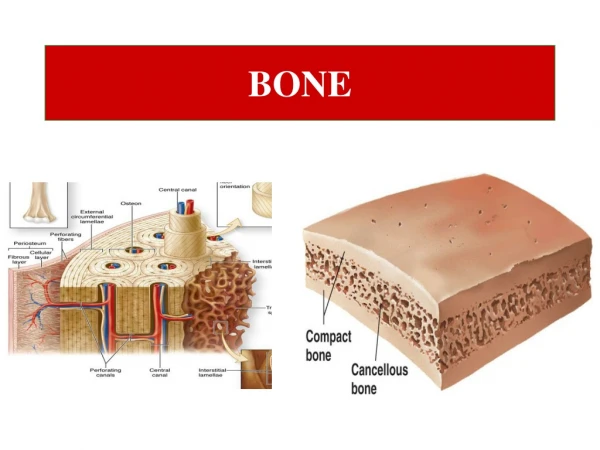
Septum of maxillary sinus The tendency for the maxillary sinus to pneumatize and form multiple lobes may give rise to the appearance of radiopaque lines extending from the floor of the sinus into the radiolucent interior. These white lines represent cortical extensions of the wall of the sinus and represent the wall of a smaller compartment within the sinus. Because these walls subdivide the sinus they are termed sinus septa or septum (singular).
Zygomatic bone Zygomatic process of Maxilla b.
Maxilla bone Zygomatic process of maxilla Zygomatic bone Zygomatic arch Zygomatic process of temporal b. Temporal bone
Zygomatic process of Maxilla b. White arrows denote the zygomatic process (generally over the first molar) Black arrows delineate the lower border of the zygomatic arch
Median palatal suture The mid-palatine suture appears in this central incisor periapical projection as a dark, or radiolucent, line at the midline (white arrows). You can also see the more radiopaque inverted triangle at the top of the image that represents the anterior nasal spine.
Incisive nerve foramen The incisive foramen is the opening in the midline of the palate just posterior to the central incisors.
Incisive foramen 1 - Incisive Foramen 2 - Nasal septum
Coronoid process This is the thin triangular prominence off the upper part of the mandible
Coronoid process Black arrows delineate margin of coronoid process
Inferior alveolar canal The mandibular canal extends from the mandibular foramen, on the lingual aspect of the ramus, through the body of the mandible under the roots of the molar teeth.
Inferior alveolar canal The inferior alveolar canal or mandibular canal runs from the lingular area of the mandible to the mental foramen and radiographically. is outlined by thin opaque edges to the canal. Its contents are the inferior alveolar nerve, artery and vein.
External oblique ridge The external oblique ridge is a ridge of bone located along the facial of the mandible, which extends from the superior aspect of the posterior body of the mandible down to the necks of the molar teeth. It runs in the same direction as the internal oblique ridge, but is located on the facial, or external surface of the mandible To distinguish radiographically between the internal and external oblique ridges, note that the external ridge is always superior to the internal oblique ridge