Download

1 / 23
230 likes | 625 Vues
Explore the detailed anatomy of the maxilla upper jaw, including clinical notes and dentoalveolar topography. Learn about sinus maxillaris, nerve and blood supply, dental implants, and more. Essential for anesthesia, extraction, implantology, and endodontic treatment.

E N D
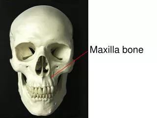
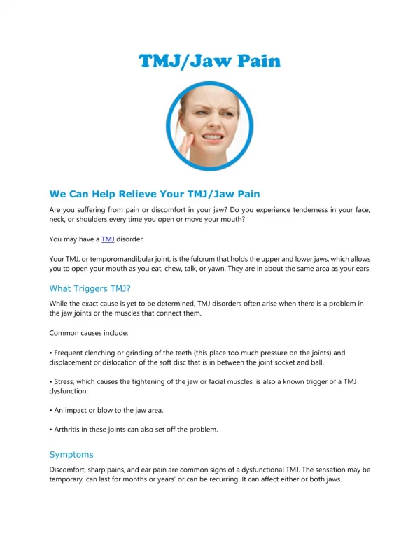
MAXILLA Upper jaw
Anatomy (repetition), widespread description • Clinical notes • Dentoalveolar topography: -transverse asymmetry of alveolus - rate of spongy and compact bone - the relationship of root to neighouring struct. • Nerve and blood supply (repetition)
Sinus maxillaris-foramina accessoria Below pr. uncinatus form the medial wall of sinus the collagenous tissue = fontanella ant. et post.in whichfor. accessoria may be occure
25-30% • Solitary or multiple • Congenital or secondary to disease process
Sinus maxillaris – decrease of floor The toothed jaw The edentulous jaw Variable layer of spongy bone between sinus and roots of teeth CAVE! By the maxillary sinus lift (augmentation) before instalation of implants
Sinus maxillaris - septa Primary: arising from the development of the maxilla Secondary: arising from the pneumatization of the sinuss floor following tooth septa 25% - 35%
CAVE! • The separately maxillary sinus puncture • Dental implants
Corpus maxillae - facies ant. (fossa canina) Caldwell-Luc antrostomy
Corpus maxillae - facies post.(tuber maxillae) • CAVE! • Alveolar foramens: a.,v.,n. alveolaris sup. post. - local anesthesia • Thin bone → during molar teeth extraction can occur maxillary tuberosity fractures
Corpus maxillae - facies orbitalis - canalis infraorbitalis CAVE ! Maxillary sinus disease can lead to dehiscence of the orbital floor → secondary neuralgia of trigeminal nerve
Palatum – zones of mucous membrane 1 – the marginal zone 2 – the incisive papilla 3 – the adipose zone 4 – the zone of the palatine seam, mucoperiosteum 5 – the glandular zone 6 – the soft palate
A H Palatum: A and H line A line localized on the line between hard and soft palate H lineline between mobile and immobile parts of the soft palate
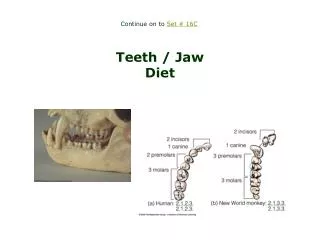
Dentoalveolar topography Important foranesthesia, extraction, injury, implantology, endodontic treatment ... The transverse asymmetry of alveolus The rate of the spongy and the compact bone The relationship the roots the upper jaw to neighbouring structures
1. The transverse asymmetry of alveolus • The dental and skeletal arch are asymmetric ! • Roots of the teeth: 1-5 eccentric směrem vestibulárním 6-7 in alveolar process axis
2. The rate of the spongy and the compact bone • The layer of compact bone is thinner than in the lower jaw • Roots of the 1-5 are surrounded by the compact bone. Posterior there are variable layer of retroalveolar spongybone. The width of the alveolus depend on the arching palate • Roots of the molars are surrounded by thin layer of the compact bone (except infrazygomatic crest)
Incisivi, canini, premolars Molars Compact bone and variable thickness of spongy bone lingually Compact bone only
3. The relationship the roots the upper jaw to neighbouring structures • Nasal cavity • Infraorbital foramen • Maxillary sinus
Nasal cavity Infraorbital foramen • Variable layer of spongy bone between nasal cavity and roots of incisivi • Root of 3 localized between nasal cavity and sinus maxillaris CAVE! Radices 1,2: periapical inflammatory may led to abscess of the floor of nasal cavity Radix 3: relation to a.,v., n. infraorbitalis and - possible trombophebitis of cavernous sinus
Maxillary sinus Variable layer of spongy bone between maxillary sinus and roots of posterior teeth CAVE! • Periapical inflammation developing at the root apices of maxillary molars and premolars are very close to the floor of the maxillary sinus - sinusitis or empyema • Potential oro-antral communication by the extraction
Trigeminal nerve Maxillar nerve - infraorbital nerve ant. sup. alv. nerve middle sup. alv. nerve post. sup. alv. nerve
Maxillary artery Post. sup. alveolar a. Infraorbital a. ant. sup. alveolar a.