Download
1 / 39
520 likes | 1.98k Vues
FRACTURES OF MAXILLA AND MANDIBLE. By DR.CHAMPA SUSHEL MBBS- FCPS ASSISTANT PROFESSOR SURGICAL UNIT -4. Etiology. Maxillofacial fractures result from either blunt or penetrating trauma.

E N D
FRACTURES OF MAXILLA AND MANDIBLE By DR.CHAMPA SUSHEL MBBS- FCPS ASSISTANT PROFESSOR SURGICAL UNIT -4
Etiology • Maxillofacial fractures result from either blunt or penetrating trauma. • @60% of patients with severe facial trauma have multisystem trauma and the potential for airway compromise. • 20-50% concurrent brain injury. • 1-4% cervical spine injuries. • Blindness occurs in 0.5-3%
Etiology • 25% of women with facial trauma are victims of domestic violence. • 25% of patients with severe facial trauma will develop Post Traumatic Stress Disorder
Emergency ManagementAirway Control • Control airway: • Chin lift. • Jaw thrust. • Oropharyngeal suctioning. • Manually move the tongue forward. • Maintain cervical immobilization
Emergency ManagementIntubation Considerations • Avoid nasotracheal intubation • Consider fiberoptic intubation if available. • Alternatives include percutaneoustranstracheal ventilation and retrograde intubation. • Be prepared for cricothyroidotomy.
Emergency ManagementHemorrhage Control • Maxillofacial bleeding: • Direct pressure. • Avoid blind clamping in wounds. • Nasal bleeding: • Direct pressure. • Anterior and posterior packing. • Pharyngeal bleeding: • Packing of the pharynx around ET tube.
History • Obtain a history from the patient /witnesses • AMPLE history • Specific Questions: • Was there loss of conscious? If so, how long? • How is your vision? • Hearing problems?
History • Specific Questions: • Is there pain with eye movement? • Are there areas of numbness or tingling on your face? • Is the patient able to bite down without any pain? • Is there pain with moving the jaw?
Physical Examination • Inspection of the face for asymmetry. • Inspect open wounds for foreign bodies. • Palpate the entire face. • Supraorbital and Infraorbital rim • Zygomatic-frontal suture • Zygomatic arches
Physical Examination • Inspect the nose for asymmetry, telecanthus, widening of the nasal bridge. • Inspect nasal septum for septal hematoma, CSF or blood. • Palpate nose for crepitus, deformity and subcutaneous air. • Palpate the zygoma along its arch and its articulations with the maxilla, frontal and temporal bone.
Physical Examination • Check facial stability. • Inspect the teeth for malocclusions, bleeding and step-off. • Intraoral examination: • Manipulation of each tooth. • Check for lacerations. • Stress the mandible. • Tongue blade test. • Palpate the mandible for tenderness, swelling and step-off.
Physical Examination • Check visual acuity. • Check pupils for roundness and reactivity. • Examine the eyelids for lacerations. • Test extra ocular muscles. • Palpate around the entire orbits..
Physical Examination • Examine the cornea for abrasions and lacerations. • Examine the anterior chamber for blood or hyphema. • Perform fundoscopic exam and examine the posterior chamber and the retina.
Physical Examination • Examine and palpate the exterior ears. • Examine the ear canals. • Check nuero distributions of the supraorbital, infraorbital, inferior alveolar and mental nerves.
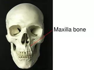
Maxillary Fractures • High energy injuries. • Impact 100 times the force of gravity is required . • Patients often have significant multisystem trauma. • Classified as LeFort fractures.
Maxillary FracturesLeFort I • Definition: • Horizontal fracture of the maxilla at the level of the nasal fossa. • Allows motion of the maxilla while the nasal bridge remains stable.
Maxillary FracturesLeFort I • Clinical findings: • Facial edema • Malocclusion of the teeth • Motion of the maxilla while the nasal bridge remains stable
Maxillary FracturesLeFort I • Radiographic findings: • Fracture line which involves • Nasal aperture • Inferior maxilla • Lateral wall of maxilla • CT of the face and head • coronal cuts • 3-D reconstruction
Maxillary FracturesLeFort II • Definition: • Pyramidal fracture • Maxilla • Nasal bones • Medial aspect of the orbits
Maxillary FracturesLeFort II • Clinical findings: • Marked facial edema • Nasal flattening • Traumatic telecanthus • Epistaxis or CSF rhinorrhea • Movement of the upper jaw and the nose.
Maxillary FracturesLeFort II • Radiographic imaging: • Fracture involves: • Nasal bones • Medial orbit • Maxillary sinus • Frontal process of the maxilla • CT of the face and head
Maxillary FracturesLeFort III • Definition: • Fractures through: • Maxilla • Zygoma • Nasal bones • Ethmoid bones • Base of the skull
Maxillary FracturesLeFort III • Clinical findings: • Dish faced deformity • Epistaxis and CSF rhinorrhea • Motion of the maxilla, nasal bones and zygoma • Severe airway obstruction
Maxillary FracturesLeFort III • Radiographic imaging: • Fractures through: • Zygomaticfrontal suture • Zygoma • Medial orbital wall • Nasal bone • CT Face and the Head
Maxillary FracturesTreatment • Secure airway • Control Bleeding • Head elevation 40-60 degrees • Consult with maxillofacial surgeon • Consider antibiotics • Admission
Mandible FracturesPathophysiology • Mandibular fractures are the third most common facial fracture. • Assaults and falls on the chin account for most of the injuries. • Multiple fractures are seen in greater then 50%. • Associated Cervical spine injuries – 0.2-6%.
Mandible FracturesClinical findings • Mandibular pain. • Malocclusion of the teeth • Separation of teeth with intraoral bleeding • Inability to fully open mouth. • Preauricular pain with biting. • Positive tongue blade test.
Mandible Fractures • Radiographs: • Panoramic view • Plain view: PA and Lateral view
Mandibular FracturesTreatment • Nondisplaced fractures: • Analgesics • Soft diet • oral surgery referral in 1-2 days • Displaced fractures, open fractures and fractures with associated dental trauma • Urgent oral surgery consultation • All fractures should be treated with antibiotics and tetanus prophylaxis.
Mandibular Dislocation • Causes of mandibular dislocation are: • Blunt trauma • Excessive mouth opening • Risk factors: • Weakness of the temporal mandibular ligament • Over stretched joint capsule • Shallow articular eminence • Neurologic diseases
Mandibular Dislocation • The mandible can be dislocated: • Anterior 70% • Posterior • Lateral • Superior • Dislocations are mostly bilateral.
Mandibular Dislocation • Clinical features: • Inability to close mouth • Pain • Facial swelling • Physical exam: • Palpable depression • Jaw will deviate away • Jaw displaced anterior
Mandibular Dislocation • Diagnosis: • History & Physical exam • X-rays • CT
Mandibular Dislocation • Treatment: • Muscle relaxant • Analgesic • Closed reduction in the emergency room
Mandibular Dislocation • Treatment: • Oral surgeon consultation: • Open dislocations • Superior, posterior or lateral dislocations • Non-reducible dislocations • Dislocations associated with fractures