Download

1 / 59
650 likes | 1.21k Vues
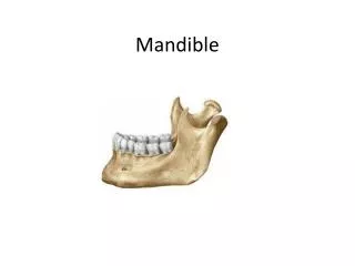
ANATOMY AND FRACTURES OF THE MANDIBLE. ANATOMY. Mandible interfaces with skull base via the TMJ and is held in position by the muscles of mastication. Anatomic units of the mandible. Muscles of the mandible – Posterior group. Muscles of the mandible – Anterior group. Muscles of Mastication.

E N D
ANATOMY • Mandible interfaces with skull base via the TMJ and is held in position by the muscles of mastication
Muscles of Mastication • OUTER SURFACE
Muscles of Mastication • INNER SURFACE
Muscles of Mastication • 4 muscles of mastication • Masseter • Temporalis • Medial pterygoid • Lateral pterygoid • Supplied by V3, testament to same embryologic origin as the mandible from the 1st branchial arch
Masseter • Divided into 3 heads • Superficial: • largest head • Arises anterior 2/3rds of the lower border of the zygomatic arch • Wide insertion to angle, forwards along lower border and upwards to lower part of ramus • Intermediate: • Middle 1/3 of the arch • Deep: • Deep surface of the arch • Action: elevator and drawing forward the angle
Masseter • Intermediate and deep fuse and pass vertically downwards to fuse with ramus • Nerve and artery divide muscle incompletely into 3 parts • Masseteric nerve (Br of anterior division of V3) runs between deep and intermediate • Br of superficial temporal and transverse facial runs between superficial and intermediate
Temporalis • Arises temporal fossa between inferior temporal line and infratemporal crest • Inserts at posterior border of the coronoid process and ascending ramus • Upper and anterior fibres elevate the mandible • Posterior fibres (horizontal) retract the mandible (only muscles that do so)
Medial pterygoid • 2 heads: • Deep: • Larger • Medial surface of the lateral pterygoid plate and the fossa between 2 plates • Superficial : • Tuberosity of the maxilla and pyramidal process of palatine bones • Insert lower and posterior part of angle (with masseter) • Action: upwards and forwards and medially
Lateral pterygoid • 2 heads: • Superior: • Infratemporal fossa • Inferior: • Lateral surface of the lateral pterygoid • Fuse into a short thick tendon that inserts into pterygoid fovea • the upper fibres passing into articular disc and anterior part of the capsule • Action: side-to-side plus only muscle to open jaw
Temporomandibular Joint • Articulation • Synovial joint between the condyle of the mandible and the mandibular fossa in the squamous part of the temporal bone • Both bone surfaces covered with layer of fibrocartilage identical to the disc • No hyaline cartilage, therefore an atypical joint
Temporomandibular Joint • Unique feature of the TMJs is the articular disc. • Composed of fibrocartilaganeous tissue • Divides each joint into 2: • Inferior compartment • Superior compartment
Temporomandibular Joint • Inferior compartment • Allows for pure rotation of the condylar head, • corresponds to the first 20 mm or so of the opening of the mouth. (opening and closing movements) • Superior compartment • involved in translational movements • sliding the lower jaw forward or side to side
Temporomandibular Joint • Atypical synovial joint separated into upper and lower cavities by a fibrocartilaginous disc • No hyaline cartilage • Capsule attached high on neck of mandible around articular margin, then to transverse prominence or articular tubercle and as far posteriorly as squamotympanic fissure • Fibrocartilage attached around periphery to capsule • Anteriorly near head of mandible, so mobile • Posteriorly near temporal bone, so more fixed • Thinner in middle than periphery, crinkled fibres to allow movement and contouring • Lateral TM ligament is a stout fibrous band passing from zygomatic arch to posterior border of neck and ramus, blending with capsule • Tightens with movements away from rest • Sphenomandibular ligament runs between sphenoid spine and lingula of mandible • Remains constant tension through range of motion as the lingula is the axis of rotation of the mandible • Sensation supplied by auriculotemporal nerve with some supply from nerve to masseter (Hiltons law)
TMJ Ligaments • 3 ligaments associated with the TMJ: • 1) Temporomandibular ligament (Major) • is really the thickened lateral portion of the capsule, and it has two parts: • an outer oblique portion (OOP) and an inner horizontal portion (IHP) • Lower border of zygomatic arch to posterior border of the neck and ramus
TMJ Ligaments • 2) stylomandibular ligament (minor) • separates the infratemporal region from the parotid region • runs from the styloid process to the angle of the mandible • 3) Sphenomandibular ligament (minor) • runs from the spine of sphenoid to the lingula of the mandible
TMJ Ligaments • The minor ligaments are important in that they define the limits of movements, • ie the farthest extent of movements of the mandible. • Not connected to joint • However, movements of the mandible made past these extents functionally allowed by the muscular attachments BUT will result in painful stimuli
Nerve Supply • Inferior alveolar nerve branch of the mandibular division of Trigeminal (V) nerve, enters the mandibular foramen and runs forward in the mandibular canal, supplying sensation to the teeth. • At the mental foramen the nerve divides into two terminal branches: • Incisive nerve: supplies the anterior teeth • mental nerve: sensation to the lower lip
Evaluation - History • Always remember ABCs of life along with secondary and tertiary survey • Mechanism of injury • MVA associated with multiple comminuted # • Fist often results in single, non - displaced # • Anterior blow to chin - bilateral condylar # • Angled blow to parasymphysis can lead to contralateral condylar or angle # • Clenched teeth can lead to alveolar process #
Physical Exam - Occlusion • Change in occlusion - determine preinjury occlusion • Posterior premature dental contact or an anterior open bite is suggestive of bilateral condylar or angle fractures • Posterior open bite is common with anterior alveolar process or parasymphyseal fractures • Unilateral open bite is suggestive of an ipsilateral angle and parasymphyseal fracture • Retrognathic occlusion is seen with condylar or angle fractures • Condylar neck # are assoc with open bite on opposite side and deviation of chin towards the side of the fx.
Angle’s classification • Class I: • Normal • Mesial buccal cusp of the upper 1st molar occludes with mesial buccal groove of the mandibular molar • Class II: • Retrocclusion, mandibular deficiency • Class III: • Prognathic occlusion, maxillary deficiency, mandibular excess
Dental classification of occlusion • Angle’s classification (1887) • Based on relationship of permanent 1st molars and to a lesser degree the permanent canines to each other
Physical Exam • Anaesthesia of the lower lip • Abnormal mandibular movement • unable to open - coronoid fx • unable to close - # of alveolus, angle or ramus • trismus • Lacerations, Haematomas, Ecchymosis • Loose teeth • swelling
Physical Exam • Multiple fractures sites are common: • 1 fracture: 50% • 2 fractures: 40% • >2 fractures: 10% • Dual patterns: • Angle contralateral body • Symphysis and bilateral condyles • 15% another facial fracture
General Principles of treatment • ABCs • Tetanus • Nutrition • Almost all can be considered open fractures as they communicate with skin or oral cavity • Reduction and fixation • Post-op monitoring for N/V, use of wire cutters • Oral care - H2O2 , irrigations, soft toothbrush
Aims of Management 1) Achieve anatomical reduction and stabilisation 2) Re-establish pre-traumatic functional occlusion 3) Restore facial contour and symmetry 4) Balance facial height and projection
Classification of Fractures • Open vs Closed • Displaced vs non-displaced • Complete vs greenstick • Linear Vs comminuted • Relationship to the teeth • Class I: teeth both sides of fracture • Class II: teeth one side of fracture • Class III: edentulous • Favourable vs unfavourable
Treatment options • No treatment • Soft diet • Maxillomandibular fixation • Open reduction - non-rigid fixation • Open reduction - rigid fixation • External pin fixation