Bone and Bone Precursors
190 likes | 424 Vues
Bone and Bone Precursors. The skeleton. Mineralized connective tissue Ligaments connecting bone to bone holds some organs in place Tendons Extension of muscle and bone. Flow chart for histogenesis of skeletal tissues. Cartilage. Dentin. Bone. Enamel. Chondroblast. Odontoblast.

Bone and Bone Precursors
E N D
Presentation Transcript
The skeleton • Mineralized connective tissue • Ligaments • connecting bone to bone • holds some organs in place • Tendons • Extension of muscle and bone
Flow chart for histogenesis of skeletal tissues Cartilage Dentin Bone Enamel Chondroblast Odontoblast Osteoblast Ameloblast Osteoprogenitors Scleroblast collagen Fibroblast Muscle cells Myoblast Mesenchymal cells
An Example: (chondrification) • Mesenchymal cells retract processes • Congregate • These cells become “Chondroblasts” • Secrete matrix about themselves • Form lacunae • Become “Chondrocytes” • Chondroblasts in middle • 2-4 cells within a lacunae – still “chondroblasts” • Result from a single cell mitotically dividing. • Secrete matrix – push away from each other – “chondrocytes” • “interstitial growth” Chondrification Center
An Example: (chondrification) • Mesenchyme cells on periphery • Differentiate to form fibroblasts that produce: • Dense irregular collagenous connective tissue (perichondrium) • Responsible for growth and maintenance of cart. • Perichondrium - two layers • Outer fibrous layer • Inner cellular layer – mainly chondrogenic cells • Mitosis to form chondroblasts –produce matrix • “appositional growth”
An Example: (chondrification) • Picture of cart.
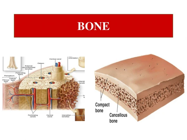
Bone • Specialized connective tissue of: • calcified bone matrix • 3 cell types • Osteocytes, in lacunae • osteoblasts make new matrix and maintain old matrix • osteoclasts: multinucleated giant cells phagocytose bone matrix in remodeling bone • Viewed as ground bone sections or decalcified and embedded paraffin sections • Endosteum and periosteum
Bone Functions • Support soft tissues • Locomotion • Protect vital organs (skull, ribs, vertebrae) • Bone marrow (hematopoiesis) • Process of differentiation of various cells from stem cells • Reservoir of calcium and phosphate
Bone Matrix • Synthesized and maintained by osteoblasts and osteocytes • 50% of dry wgt is inorganic: calcium and phosphate • Hydroxyapatite crystals • Organic part is 95% type I collagen with amorphous ground substance (glycoproteins)
Compact lamellar bone Membrane Bone
Compact lamellar bone