Download
1 / 49
580 likes | 1.14k Vues
DENTAL GROSS ANATOMY CASE 3 INFRATEMPORAL FOSSA & TEMPOROMANDIBULAR JOINT. HISTORY Sherry Goldsmith was involved in a two-car collision and suffered severe facial injuries. EXAMINATION In the ER she was found to have the following injuries:

E N D
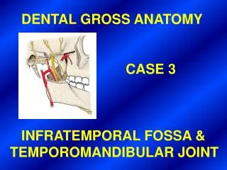
DENTAL GROSS ANATOMY CASE 3 INFRATEMPORAL FOSSA & TEMPOROMANDIBULAR JOINT
HISTORY • Sherry Goldsmith was involved in a two-car collision and suffered severe facial • injuries. • EXAMINATION • In the ER she was found to have the following injuries: • The ramus of the mandible was shattered on the left side and displaced • medially into the infratemporalfossa. • The condyloid and coronoid processes were broken off on the left side. • The TMJ was driven medially breaking off the spine of the sphenoid. • A sliver of the windshield passed deeply into the infratemporalfossa, • reaching beyond the level of the infratemporal crest. • A large hematoma was found within the fossa shrouding the other contents. • After debridement of the wounds, an MRI was performed to determine the • total amount of the injuries.
POSTOPERATIVE OUTCOME • After a lengthy hospitalization and numerous surgeries, Ms. Goldsmith • was found to have the following neural and neuromuscular disorders: • Ipsilateral loss of taste sensations on the anterior part of the tongue. • Ipsilateral loss of general sensations on the anterior part of the tongue. • When the patient opens her jaw, the mandible deviates toward the side • of the injury. • Ipsilateralcutaneous anesthesia involving a strip of skin extending from • the lower lip and proceeding anterior to the ear and superior to the scalp. • Ipsilateral anesthesia of the lingual gingiva (mandibular region), floor of • the mouth and mandibular teeth. • 6. There was a reduction of the volume of saliva.
1. What are the bony boundaries of the infratemporal fossa?
BOUNDARIES OF THE INFRATEMPORAL FOSSA Ramus of mandible (lateral wall)
BOUNDARIES OF THE INFRATEMPORAL FOSSA Temporal fossa Infratemporal crest of greater wing of sphenoid Infratemporal surface of greater wing of sphenoid (roof) Infratemporal surface of maxilla (anterior wall) Styloid process (posterior wall) Lateral pterygoid plate (medial wall)
Name the six, usually expected, • contents of the infratemporal fossa. • Muscles of mastication • (except masseter) • Pterygoid plexus of veins • 1st and 2nd parts of maxillary a. • (mandibular and pterygoid parts) • Mandibular division of V (V3) • Otic ganglion • Chorda tympani n.
CONTENTS OF INFRATEMPORAL FOSSA Mandibular n. (V3) Lateral pterygoid m. Chorda tympani n. Medial pterygoid m.
CONTENTS OF INFRATEMPORAL FOSSA (MEDIAL VIEW) V3 Otic ganglion Chorda tympani n. Maxillary a. Medial pterygoid m.
CONTENTS OF INFRATEMPORAL FOSSA Superficial temporal v. Pterygoid plexus Maxillary vv. Retromandibular v.
Discuss the specific attachments • and actions of the muscles of • mastication.
Temporalis fascia Temporalis m. Zygomatic arch Deep part (masseter m.) Superficial part (masseter m.)
Temporalis m. Insertion of temporalis m. into coronoid process and anterior border of ramus of mandible Insertion of masseter m. into lateral surface of ramus of mandible
Articular disc of TMJ Superior and inferior heads of lateral pterygoid m. Superficial and deep heads of medial pterygoid m. Pterygoid fovea of neck of mandble Angle of mandible
Explain the peculiar deviation • of the intact mandible.
Lateral pterygoid plate V3 injured on this side Lateral pterygoid m. Medial pterygoid m. Deviation of mandible to paralyzed side
5. Name the foramen through which the mandibular division of the trigeminal nerve (V3) passes into the infratemporal fossa. In which boundary of the infratemporal fossa is this foramen located? Does V3 supply any muscles other than the muscles of mastication?
Foramen ovale (V3) in roof of infratemporal fossa
V3 SUPPLIES ALL MUSCLES DERIVED FROM PHARYNGEAL ARCH 1 Temporalis Masseter Mylohyoid & ant. belly of digastric NOT SHOWN Medial & lateral ptergoids NOT SHOWN Tensor palati & tensor tympani
Temporalis fascia and m. Anterior division (V3) (mostly motor) Posterior and anterior deep temporal nn. Posterior division (V3) (mostly sensory) Foramen ovale Masseteric n. Lateral pterygoid n. and m. Auriculotemporal n. Chorda tympani n. Buccal n. (sensory) Lingual n. Inferior alveolar n. (cut) Mylohyoid n. (motor) Mylohyoid m. (cut) Inferior alveolar n. (cut) Digastric m. (anterior belly)
Motor root Sensory root Mandibular n. (V3) Tensor (veli) palatini m. and n. Tensor tympani m. and n. Medial pterygoid m. and n.
Explain the cutaneous loss • demonstrated by the patient.
V3 Mental n. Buccal n. Auriculotemporal n.
Explain the loss of taste on the • anterior part of the tongue.
Geniculate ganglion (of VII) (sensory neurons) Chorda tympani n. (br. of VII) supplies taste buds on anterior 2/3 of tongue
Explain the decreased volume • of saliva.
V3 Lesser petrosal n. Otic ganglion IX Auriculotemporal n. Inferior salivatory nucleus Parotid gland Tympanic plexus Tympanic n. Presynaptic parasympathetic fibers Postsynaptic parasympathetic fibers
Lingual n. Chorda tympani n. VII Superior salivatory nucleus Submandibular ganglion Sublingual and submandibular glands Presynaptic parasympathetic fibers Postsynaptic parasympatheti fibers
9. a. Identify the branch of the maxillary artery which enters the middle cranial fossa. From what part of the maxillary a. does it arise? b. Discuss the pathway by which this artery enters the middle cranial fossa.
Anterior and posterior deep temporal aa. Lateral pterygoid a. and m. Masseteric a. Middle meningeal a. Maxillary a. Infraorbital a. Posterior superior alveolar a. Inferior alveolar a. Buccal a. Medial pterygoid a. and m. MAXILLARY A. BRANCHES OF 1ST (MANDIBULAR) PART BRANCHES OF 2ND (PTERYGOID) PART BRANCHES OF 3RD (PTERYGOPALATINE) PART
Middle meningeal a. (entering foramen spinosum) Auriculotemporal n.
9. c. What does this artery supply? d. Name the condition which results from tearing this artery within the cranial cavity. What will be the consequence if this injury is not repaired immediately?
Calvaria Dura mater (Does not supply the brain) Middle meningeal a.
Through what fissure does the maxillary • artery extend medially out of the • infratemporal fossa?
Pterygomaxillary fissure (conducts maxillary a. into pterygopalatine fossa)
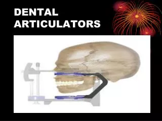
11. a. What type of joint is the TMJ? Be specific. b. What structure lies inside the TMJ and what kinds of movement occur in each part of the joint?
TMJ IS A MODIFIED HINGE TYPE OF SYNOVIAL JOINT Lower joint compartment (hinge action) Upper joint compartment (gliding action) Articular disc (avascular fibrous tissue) Articular tubercle Joint capsule
12. Describe the movements of the head of the mandible when the mouth is opened widely.
JAW CLOSED JAW WIDELY OPENED
Name the parts of the mandibular • fossa and their boundaries.
Articular tubercle Mandibular fossa (articular part formed by squamous temporal) Postglenoid tubercle Mandibular fossa (nonarticular part formed by tympanic temporal)
14. What bony structure offers resistance to medial displacement of the head of the mandible?
Middle cranial fossa Articular disc Spine of sphenoid
15. Discuss the intrinsic and extrinsic • ligaments of the TMJ and the specific • function of the lateral thickening of the • fibrous capsule. • Name the arterial supply and nerve • supply of the TMJ.
Joint capsule Lateral (temporomandibular) ligament Styloid process Stylomandibular ligament . Auriculotemporaln. Maxillary a. Inferior alveolar n. Lingual n. Sphenomandibular ligament INTRINSIC LIGAMENT EXTRINSIC LIGAMENTS Mylohyoid a. and n.
Explain • (a) the movement resulting in the most • common displacement of the TMJ • (b) the “clicking” sound produced by • chronic dislocation of this joint.