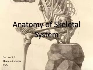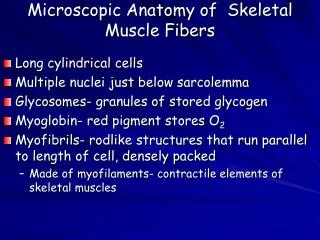
Anatomy of Skeletal Elements
780 likes | 1.78k Vues
Anatomy of Skeletal Elements. Bone Surface Markings. Foramen = opening (arteries, nerves) Fossa = shallow depression Sulcus = shallow groove (artery or nerve) Canal = longer, tubelike opening Fissure = narrow, cleftlike opening Notch = indentation at the end of a bone

Anatomy of Skeletal Elements
E N D
Presentation Transcript
Bone Surface Markings • Foramen = opening (arteries, nerves) • Fossa = shallow depression • Sulcus = shallow groove (artery or nerve) • Canal = longer, tubelike opening • Fissure = narrow, cleftlike opening • Notch = indentation at the end of a bone • Meatus = type of canal • Condyle = large, round protuberance, attachment of muscles • Epicondyle = above or upon a condyle • Facet = smooth flat articular surface • Trochanter = very large projection • Tuberosity = large, rounded, roughened projection • Tubercle = rounded eminence/elevation • Crest = roughened border or ridge • Spine = sharply pointed projection
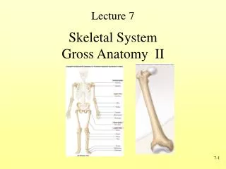
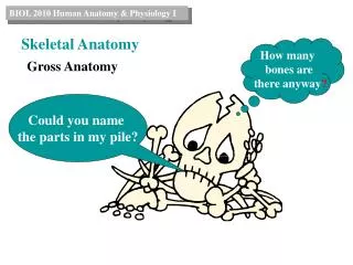
Skeletal system includes • Axial division • Skull and associated bones • Auditory ossicles • Hyoid bones • Vertebral column • Thoracic cage • Ribs sternum • Appendicular division • -Pectoral girdle • -Pelvic girdle
The Axial Skeleton • Axial division • Skull and associated bones • Auditory ossicles • Hyoid bones • Vertebral column • Thoracic cage • Ribs sternum
Sutures • Immovable joints (synarthrotic, fibrous joints) • Form boundaries between skull bones • Five sutures • Coronal • Sagittal • Lambdoid • Squamous • Frontonasal
The Adult Skull • skull = 22 bones • cranium = 8 bones: frontal, occipital, 2 temporals, 2 parietals, sphenoid and • ethmoid • facial bones = 14 bones: nasals, maxillae, zygomatics, mandible, lacrimals, • palatines, inferior nasal conchae, vomer. • skull forms a larger cranial cavity • -also forms the nasal cavity, the orbits, paranasal sinuses • mandible and auditory ossicles are the only movable skull bones • cranial bones also: attach to membranes called meninges • -stabilize positions of the brain, blood vessels • -outer surface provides large areas for muscle attachment that • move the head or provide facial expressions
Frontal bone • Forms the forehead • Roof of the orbit • articulates with parietal, sphenoid, lacrimal, nasal, ethmoid, zygomatic and maxilla • superior and lateral to glabellar region – frontal sinuses • inferior portion – supraorbital ridges with supraorbital notch (supraorbital nerve and artery)
Parietal bones • -Part of the superior and lateral surfaces of the cranium • -articulate with each other – sagittal suture • -articulate with occipital, frontal, • temporal and sphenoid bones
Temporal bone • -Forms wall of jugular foramen • -Petrous part: posterior portion • -Tympanic part: associated with ear canal • -Squamous part: anterior portion, fan-shaped • -zygomatic process • -forms cranial portion of the TMJ joint • -inferior to zygo. process – mandibular fossa (mandibular condyle)
Temporal bone – petrous portion • houses the inner ear • -inferior aspect – mastoid process (air spaces that communicate with the middle • ear) • -also for attachment of sternocleidomastoid muscle • -inferior to mastoid process – mastoid foramen • -anterior to mastoid process – external acoustic meatus • -inferior and medial to the MP – styloid process (muscle attachment) • -stylomastoid foramen (7th cranial)
Occipital bone • Part of the base of the skull • articulates with parietal, temporal and sphenoid • Surrounds the foramen magnum • lateral to the FM – hypoglossal canal (12th cranial) • projections = occipital condyles • Forms part of the jugular foramen
Sphenoid bone • Contributes to floor of cranium • articulates with the frontal, ethmoid, temporal zygomatic, parietal maxillary, palatine, vomer & occipital bones • Bridges cranial and facial bones • Optic canal allows passage of optic nerve
-Foramina: -superior orbital fissure – 3rd, 4th, 6th cranials -foramen ovale – 5th cranial (mandibular) -foramen rontundum – 5th cranial -foramen spinosum – middle meningeal artery -optic canal • -greater wing – posterolateral portion • -lesser wing • -Pterygoid processes – inferior to the greater wings – sites of muscle attachment • -lateral and medial plates with a • fossa located between • -body: sella turcica, tuberculum sellae, dorsum sellae, sphenoid sinuses (paranasal sinuses)
Irregularly shaped bone • Forms part of orbital wall • Forms roof of nasal cavity • articulates with: frontal, sphenoid, lacrimal and maxillary bones • connects with the vomer • two lateral masses – contain the ethmoid sinuses • projections called the superior and middle nasal conchae • two plates: perpendicular plate & the cribiform plate • Cribiform plate: perforations for olfactory nerve, midline is the crista galli • Perpendicular plate = upper part of nasal septum Ethmoid bone
14 Facial Bones Nasal (2) Maxillae (2) Zygomatic (2) Mandible (1) Lacrimal (2) Palatine (2) Inferior nasal conchae (2) Vomer (1)
Bones of the Face • Maxillae • Paired bone • Largest of facial bones • Form upper jaw • body = orbital, nasal, infratemporal and facial surfaces • body contains the maxillary sinuses
Maxillary bones: Anterior View • frontal process – articulates with frontal bone and nasal bones & forms medial orbital rim • facial surface – body of maxilla • infraorbital foramen – landmark for local • inferior to this IF – canine fossa • inferior portion of the maxilla – alveolar processes of the teeth (contains the roots of the maxillary teeth
zygomatic process: articulates with zygomatic bone • Inferior view: • palatine process – major portion of the hard palate • joined by the median palatine suture covered by the median palatine raphe (fibrous tissue) • anterior portion – incisive foramen (nasopalatine nerves and vessels)
Zygomatic Bones • Cheekbones • Lateral wall of orbit along with sphenoid • Part of zygomatic arch along with part of temporal
Palatine bones • Small, L-shaped • link between maxilla and sphenoid • Form posterior portion of hard palate • Contribute to floor of orbit • made up of horizontal plate and a vertical plate + orbital process
-midpoint of horizontal plate = nasal crest -vertical plates – forms lateral wall of nasal cavity -two foramina in each palatine bone 1. greater palatine – about 3rd molar -greater palatine nerve, landmark for administration of local 2. lesser palatine – lesser palatine nerve to soft palate
Inferior nasal concha • Located on each side of nasal septum • Increase epithelial surface • Create turbulence in inspired air • Lacrimal bones • Smallest bones in skull • Forms nasolacrimal groove leading to nasolacrimal canal • Delivers tears to nasal cavity
Palatine & Vomer • Vomer • posterior part of nasal septum • Forms inferior portion of nasal septum • Articulates with maxillae and palatines
Mandible • lower jaw • only freely movable bone of the skull • moving articulations with temporal bone
Mandible: Anterior surface • many landmarks on the body • mental protruberence – beneath the roots of the mandibular incisors • faint ridge at the midline – mandibular symphysis (fusion of right and left processes during development) • lateral to the midline – mental foramina (mental nerve and vessels into the mandibular canal)
Mandible: Anterior & Lateral surfaces • superior to the body – alveolar processes of the mandible • body is capable of elongation in children • like the maxillary alveolar processes, the alveolar processes of the mandible can become completely resorbed upon loss of teeth • ramus – superior and posterior to the body • primary area for attachment of muscles for mastication • also grows • anterior border is the coronoid process • posterior border – mandibular condyle • between in the mandibular notch or coronoid notch (landmark for local) • ramus and body joined at the external oblique line • superior to this – coronoid notch
Mandible: Inferior surface -visible are the genial tubercles – or mental spines -muscle attachment area -two fossas: 1) sublingual (sublingual salivary gl.) 2) submandibular (submandibular gl.) -divided by the mylohyoid line (mylohyoid m.) -mandibular foramen – opening of the mandibular canal -for the exit of the alveolar nerve and vessels -can be lost with alveolar process reabsorption -overhanging the foramen – lingula (attachment of sphenomandibular ligament – TMJ)
The Hyoid Bone • Suspended by stylohyoid ligaments • Consists of a body, greater horns and lesser horns • Base for muscles of the tongue and larynx
The Orbital and Nasal Complexes • Orbital complex • Bony recess that holds the eye • Seven bones • Frontal bone • Lacrimal bones • Palatine bones • Zygomatic bones • Ethmoid • Sphenoid • Maxillae
Orbital Complex: Eye socket • medial wall: frontal process, lacrimal bone and part of ethmoid • lateral wall: sphenoid, zygomatic • floor: maxillary, zygomatic • back: sphenoid + superior orbital fissure -top: frontal bone • sphenoid and frontal bones are separated by the infaorbital fissure (infraorbital & zygomatic nerves, infraorbital artery and inferior opthalmic vein) -continues on as the infraorbital sulcus -becomes the infraorbital canal -terminates on the facial surface as the infraorbital foramen (infraorbital nerve)
The Nasal Complex • Bones and cartilage that enclose the nasal cavity
Nasal bones & cavities • Nasal bones • Paired bones – forms the bridge • Articulate with frontal bone • nasion: junction between frontal and nasal bones • Nasal cavity • anterior, triangular opening: piriform aperture -lateral wall: nasal conchae (superior, middle, inferior) -superior and middle - ethmoid -inferior nasal conchae – separate bone -divided into separate cavities – nasal septum -anterior portion is nasal septal cartilage -superior portion formed by perpendicular plate -inferior portion formed by the vomer deviated nasal septum: nasal septum divides the nasal cavity into right and left halves -three components: vomer, septal cartilage & perpendicular plate of the ethmoid -deviation results in a later deflection of the septum -severe deviation may affect breathing
Paranasal Sinuses • frontal sinuses: frontal bone, separated by a septum • connects with nasal cavity – frontonasal duct • sphenoid sinuses: body of the sphenoid bone • also drain into nasal cavity • ethmoid sinuses: or ethmoid air cells, located in the lateral masses • anterior, middle and posterior sinuses • maxillary: body of the maxilla • size varies with individual and age • largest of the sinuses • close proximity to alveolar processes – periodontal tissues may be in direct contact with sinus’ mucus membranes • part of the nasal complex • Paired cavities in ethmoid, sphenoid, frontal and maxillary • Lined with mucous membranes and open into nasal cavity though openings called ostia • Resonating chambers for voice, lighten the skull • Sinusitis is inflammation of the membrane (allergy) • infection can easily spread from one sinus to the other through the nasal cavity • can also spread to other tissues • secondary sinusitis
Cranial Fossae • Depressions in cranial floor • Anterior cranial fossa • Frontal bone, ethmoid, lesser wings of sphenoid • Middle cranial fossa • Sphenoid, temporal bones, parietal bones • Posterior cranial fossa • Occipital bone, temporal bones, parietal bones