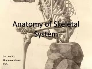
Skeletal system anatomy
Skeletal system anatomy. The skeletal system. Functions : Protection for vital organs Serves as levers for movement Storage site for minerals Site for blood formation. Parts or layers of bone: Periosteum Compact bone Bone Marrow Cartilage. Periosteum.

Skeletal system anatomy
E N D
Presentation Transcript
The skeletal system Functions : • Protection for vital organs • Serves as levers for movement • Storage site for minerals • Site for blood formation
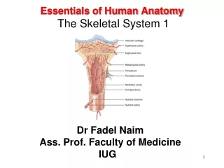
Parts or layers of bone: • Periosteum • Compact bone • Bone Marrow • Cartilage
Periosteum The fibrous sheath that covers bones. It contains the blood vessels and nerves that provide nourishment and sensation to the bone.
Compact bone • honeycombed • passages for blood vessels and nerves • bony tissue • calcium • Phosphorus
Bone Marrow • gelatinous • Yellow marrow mostly fat • Red marrow • red blood cells • white blood cells • platelets
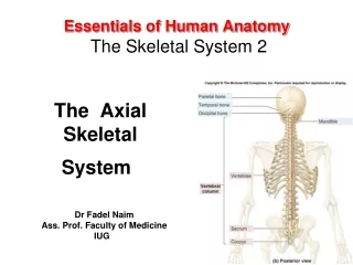
Parts of the skeletal system • Axial skeleton • Appendicular skeleton • Viesceral skeleton
Axial skeleton • vertebral column (Columnavertebralis) • Ribs (Os Costae) • Sternum (Os Sternum) • Skull (Ossa Cranii)
Columnavertebarlis • Protects spinal cord • Consist of : • Vertebrae cervicales • Vertebrae thoracales • Vertebrae lumbales • Vertebrae sacrales • Vertebrae cocygeae
Vertebrae cervicales • Involved with head and neck movement • Most flexible part of the Axial Skeleton • Seven V. Cervicales in all species
Vertebrae thoracales • Limited movement and flexibility • Located at the dorsal area of thoracic region
Vertebrae lumbales • Framework for loin area • More flexibility than thoracic but less than cervical
Vertebrae sacrales • Formed by the fusion of five vertebrae, and is conveniently described as a single, the sacrum
sternum • A median segmental bone. It consists of six to eight bony segments (sternebrae) connected by intervening cartilage in the young animal
Skull (Ossa cranii) • Divide into : • The cranial bones (Ossa cranii) inclose the brain with its membranes and vessels and the essential organs of hearing • The facial bones (Ossa facici) form the skeleton of the oral and nasal cavities and also support the pharynx, larynx and the root of the tongue
Os cranii Consist of : • The occipital bone (Os occipitale/tulangkepalabelakang) • The sphenoid bone (Os sphenoidale/tulangbaji) • The Ethmoid bone (Os ethmoidale/tulangtaji) • The parietal bones (Ossa parietalia) • The frontal bones (Ossa frontalia/tulangdahi) • The temporal bone (Os temporale/tulangpelipis)
Os facici Consist of : • The maxillae (tulangrahangatas) • The premaxillae (Ossa incisiva/tulangrahangatasmuka) • The nasal bones (Ossa nasalia/ tulanghidung) • The lacrimal bones (Ossa lacrimalia/tulang air mata) • The mandible (Mandibula/ rahangbawah)
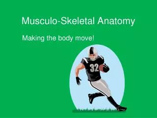
Appendicular skeleton • Locomotion • Eating • Connected to axial Skeleton by muscles &/or Bony Joints
Appendicular skeleton Divide into : • The thoracic limb (Extremitasthoracalis) • The Pelvic limb (Extremitaspelvina)
The thoracic Limb • The shoulder girdle (Cingulumextremitatisthoracalis) scapula (shoulder blade) • The arm (Brachium) humerus (arm bone) • Forearm (Antibrachium) radius and ulna • Manus (homologoue of the hand in man) carpus, metacarpus, digits (homologus with fingers in human)
Scapula 1, the acromion process; 2, scapular spine; 3, the glenoid cavity; and 4, the scapular cartilage
humerus 1, lateral tuberosity; 2, deltoid tuberosity; 3, lateral condyloid crest; 4, coronoidfossa; 5, lateral condyle; 6, medial condyle; 7, musculo-spiral groove; 8, medial tuberosity; 9, intertuberal groove; 10, articular head; 11, medial epicondyle; and 12, lateral epicondyle.
Radius and Ulna 1, distal end of humerus; 2, olecranonfossa; 3, olecranon process;, 4,radius; 5, ulna; and 6, carpal bones.
Carpus (Ossa carpi) AC, accessory carpal; C, carpal; IC, intermediate carpal; MC, metacarpal; P, phalanges; RC, radial carpal; and UC, ulnar carpal
The Pelvic limb Consist of : • The pelvic (Cingulumextremitaspelvinae) oscoxae/hip bone (Ilium, ischium, and pubis) • The thigh (femur) • The skeleton of the leg (Crus) tibia, fibula and patella • The pes (homologue of the foot in human) tarsus, metatarsus and digits
femur 1, trochanter major; 2, head of femur; 3, trochantericfossa; 4, neck of femur; 5, trochanter minor; 6, lateral supracondyloid crest; 7, supracondyloidfossa; 8, trochlea; 9, extensor fossa; 10, lateral epicondyle; 12, intercondyloidfossa; and 13, medial condyle.
Tibia and fibula 1, medial condyle, 2, lateral condyle; 3, tibia, and 4, fibula.
The viesceral skeleton • Consist of certain bones developed in the substance of some viscera or soft organs, such as : os penis of the dog, and oscordis of the ox
Classification of bones (based on the shape) • Long bones (Ossa longa) humerus, femur • Flat bones (Ossa plana) scapula, oscoxae • Short bones (Ossa brevia) carpus, tarsus • Irregular bones (Ossa irregularia) vertebral column