Download
1 / 29
290 likes | 606 Vues
Musculo-Skeletal Anatomy. Making the body move!. Goals. Important muscle groups to know Review muscle functions, types, and general anatomy In-depth look at skeletal muscle: Connective tissue covering Skeletal muscle fibers Neuromuscular junction. Three types of muscle. Skeletal Cardiac

E N D
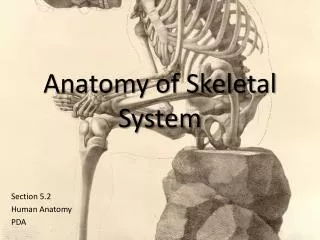
Musculo-Skeletal Anatomy Making the body move!
Goals • Important muscle groups to know • Review muscle functions, types, and general anatomy • In-depth look at skeletal muscle: • Connective tissue covering • Skeletal muscle fibers • Neuromuscular junction
Three types of muscle • Skeletal • Cardiac • Smooth
The Anterior (see handout) Anterior = “Front”
The Posterior (See handout) Posterior = “back”
Functions of Muscle • Producing movement. • Maintaining posture. • Stabilizing joints. • Generating heat.
Characteristics of Muscles (some review, some *new*!) • Muscle cells are elongated (*muscle cell = muscle fiber) • *Contraction of muscles is due to the movement of microfilaments • *All muscles share some terminology • Prefix myo refers to muscle • Prefix mys refers to muscle • Prefix sarco refers to flesh
Skeletal Muscle Characteristics • *Most are attached by tendons to bones • Cells have many nuclei • Striated – have visible banding • Voluntary – subject to conscious control • *Cells are surrounded by and bundled by connective tissue
Skeletal Muscle • Striated, long, cylindrical, multi-nucleated. • Attached to bones • Voluntarily controlled • * Range of contractile speed
Cardiac Muscle • Striated, one nucleus, branching chains of cells, intercalated discs (= dark bands) • Found only in the heart. • Involuntary contraction. • *Slow speed of contraction. • *Rhythmic contraction.
Smooth Muscle • One nucleus, no striations. • *Found along the walls of blood vessels and digestive canals. • Involuntary contraction. • *Very slow contraction.
Skeletal Muscle Attachments • What structures do muscles attach to? • Bones • Cartilages • **extensions of connective tissue coverings
Connective tissue coverings • Layers of connective tissue enclose and separate all parts of a skeletal muscle • Fascia • Epimysium • Perimysium • Endomysium Friends Enjoy Protecting Each other
Fascia • layers of connective tissue that separate individual skeletal muscles from each other • hold muscles in position • May extend beyond muscle to form: • Tendon – cord-like structure • Aponeuroses – sheet-like structure
Connective Tissue Wrappings of Skeletal Muscle • Fascia – on the outside of the epimysium • Epimysium – covers the entire skeletal muscle Figure 6.1
Connective Tissue Wrappings of Skeletal Muscle • Perimysium – around a fascicle (bundle) of fibers • Endomysium – around single muscle fiber Figure 6.1
How does the structure of the coverings arrangement allow the muscles to function? • Structure: layers of connective tissue enclose and separate all parts of a skeletal muscle • Function: • allows parts to move somewhat independently • Blood vessels and nerves pass through the layers allows for blood/nutrient supply and stimulation
Review • Produce movement • Maintain posture • Stabilize joints • Generate heat • Connective tissue coverings ?
Overall structure of muscle • Muscle • Fascicles (bundles) • Muscle fibers (cells) • Myofibrils • Thick and thin filaments
Muscle fibers • Definition: single cell that contracts in response to stimulation, relaxes when stimulation ends • Thin, elongated cylinder that could extend full length of muscle
Muscle fibers • Cells are multinucleate • Nuclei are just beneath the sarcolemma
Microscopic Anatomy of Skeletal Muscle • Sarcolemma – specialized plasma membrane • Sarcoplasmic reticulum – specialized smooth endoplasmic reticulum Figure 6.3a
Microscopic Anatomy of Skeletal Muscle • Myofibril • Bundles of myofilaments • Myofibrils are aligned to give distinct bands • I band = light band • A band = dark band • Bands make muscle look STRIATED Figure 6.3b
Microscopic Anatomy of Skeletal Muscle • Sarcomere • Contractile unit of a muscle fiber • From Z disc to Z disc Figure 6.3b
Microscopic Anatomy of Skeletal Muscle • sarcomere organization (Section of myofibrils) • Thick filaments = myosin filaments = dark band • Composed of the protein myosin • Has ATPase enzymes (for contraction) Figure 6.3c
Microscopic Anatomy of Skeletal Muscle • Organization of the sarcomere • Thin filaments = actin filaments = light band • Composed of the protein actin Figure 6.3c
Microscopic Anatomy of Skeletal Muscle • Myosin filaments have heads (extensions, or cross bridges) • Myosin and actin overlap somewhat Figure 6.3d
Microscopic Anatomy of Skeletal Muscle • At rest, there is a bare zone that lacks actin filaments • Sarcoplasmic reticulum (SR) – for storage of calcium Figure 6.3d
Modeling activity • In pairs, using the available materials, create a model of a myofibril segment between 2 M lines. • Include/indicate sarcomere, actin, myosin, 2 M lines, I band, and A band