Download
1 / 21
210 likes | 228 Vues
Cell Communication. Communication Methods. Cell-to-cell contact Local signaling Long distance signaling. Plasma membranes. Plasmodesmata between plant cells. Gap junctions between animal cells. Cell-to-Cell Communications. Cell junctions directly connect the cytoplasm of adjacent cells

E N D
Communication Methods • Cell-to-cell contact • Local signaling • Long distance signaling
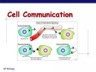
Plasma membranes Plasmodesmata between plant cells Gap junctions between animal cells Cell-to-Cell Communications • Cell junctions directly connect the cytoplasm of adjacent cells • Ex: cardiac cells for rhythmicity • Surface receptors can give/send information • Ex: specific immune response
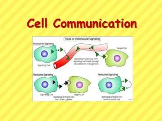
Local signaling Target cell Electrical signal along nerve cell triggers release of neurotransmitter Neurotransmitter diffuses acrosssynapse Secretory vesicle Local regulator diffuses through extracellular fluid Target cell is stimulated (b) Synaptic signaling. A nerve cell releases neurotransmitter molecules into a synapse, stimulating the target cell. (a) Paracrine signaling. A secreting cell acts on nearby target cells by discharging molecules of a local regulator (a growth factor, for example) into the extracellular fluid. Local Signaling • Adjacent cells are signaled. • Chemical messengers released • Ex: Neurotransmitters via neurons
Long-distance signaling Blood vessel Endocrine cell Hormone travels in bloodstream to target cells Target cell (c) Hormonal signaling. Specialized endocrine cells secrete hormones into body fluids, often the blood. Hormones may reach virtually all body cells. Figure 11.4 C Long Distance Signaling • Use of hormones • Both plants and animals use hormones (e.g. Insulin) • Can affect many cells in Other parts of the body • Protein or Steroid types
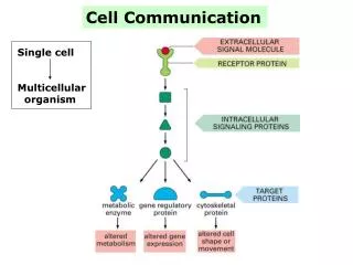
How Do Cells Communicate? • Signal Transduction Pathways • Convert signals on a cell’s surface into cellular responses • Are similar in microbes and mammals, suggesting an early origin
EXTRACELLULAR FLUID CYTOPLASM Plasma membrane 1 2 3 Reception Transduction Response Receptor Activation of cellular response Relay molecules in a signal transduction pathway Signal molecule Figure 11.5 3 Phases of Signal Transduction
Step One - Reception • Reception occurs when a signal molecule (ligand) binds to a receptor protein. • Receptor protein is on the cell surface • Ligand and receptor have a unique bonding
Step Two - Transduction • Signal initiated by conformational change of receptor protein • Signal is turned into a cellular response. • Signaling cascades relay signals to target • Multistep pathways can amplify a signal • Second messengers involved
Signal molecule A relay molecule activates protein kinase 1. Receptor Activated relay molecule 4 1 3 5 2 Inactive protein kinase 1 Active protein kinase 1 transfers a phosphate from ATP to an inactive molecule of protein kinase 2, thus activating this second kinase. Active protein kinase 1 Active protein kinase 2 then catalyzes the phos- phorylation (and activation) of protein kinase 3. Inactive protein kinase 2 ATP Phosphorylation cascade P Active protein kinase 2 ADP PP P i Enzymes called protein phosphatases (PP) catalyze the removal of the phosphate groups from the proteins, making them inactive and available for reuse. Inactive protein kinase 3 Finally, active protein kinase 3 phosphorylates a protein (pink) that brings about the cell’s response to the signal. ATP P ADP Active protein kinase 3 PP P i Inactive protein ATP P ADP Active protein Cellular response PP P i • A phosphorylation cascade Figure 11.8
First messenger (signal molecule such as epinephrine) Adenylyl cyclase G protein GTP G-protein-linked receptor ATP cAMP Protein kinase A Cellular responses Cyclic AMP example…
3 2 1 4 6 5 A signal molecule binds to a receptor, leading to activation of phospholipase C. DAG functions as a second messenger in other pathways. Phospholipase C cleaves a plasma membrane phospholipid called PIP2 into DAG and IP3. EXTRA- CELLULAR FLUID Signal molecule (first messenger) G protein DAG GTP PIP2 G-protein-linked receptor Phospholipase C IP3 (second messenger) IP3-gated calcium channel Endoplasmic reticulum (ER) Various proteins activated Cellularresponse Ca2+ Ca2+ (second messenger) The calcium ions activate the next protein in one or more signaling pathways. IP3 quickly diffuses through the cytosol and binds to an IP3– gated calcium channel in the ER membrane, causing it to open. Calcium ions flow out of the ER (down their con- centration gradient), raising the Ca2+ level in the cytosol. Ex: Inositol P3 and calcium
Step Three - Response • Cell signaling leads to regulation of cytoplasmic activities or transcription • Signaling pathways regulate a variety of cellular activities
Growth factor Reception Receptor Phosphorylation cascade Transduction CYTOPLASM Inactive transcription factor Active transcription factor Response P DNA Gene Figure 11.14 mRNA NUCLEUS Pathways can also regulate genes by activating transcription factors that turn genes on or off
Types of Receptors • There are three main types of plasma membrane receptors: • G-protein-linked • Tyrosine kinases • Ion channel
Inactivate enzyme ActivatedReceptor G-protein-linked Receptor Signal molecule Plasma Membrane GDP G-protein(inactive) GTP GDP CYTOPLASM Enzyme Activated enzyme GTP GDP Pi Cellular response G-protein-linked receptors • Very common, diverse functions • Only results in single pathway response
Signal-binding site Signalmolecule Signal molecule Helix in the Membrane Tyr Tyr Tyr Tyr Tyrosines Tyr Tyr Tyr Tyr Tyr Tyr Tyr Tyr Receptor tyrosinekinase proteins(inactive monomers) Dimer CYTOPLASM Figure 11.7 Activatedrelay proteins Cellularresponse 1 Tyr Tyr Tyr Tyr Tyr Tyr P P Tyr P Tyr Tyr Tyr Tyr P Tyr Tyr Tyr P P P Tyr Tyr Tyr Tyr Tyr P Tyr Tyr Tyr Cellularresponse 2 P P P Tyr Tyr P 6 ATP 6 ADP Activated tyrosine- kinase regions (unphosphorylated dimer) Fully activated receptor tyrosine-kinase (phosphorylated dimer) Inactiverelay proteins Receptor tyrosine kinases • Multiple pathway response • Regulates/coordinates many cell functions
Gate closed Signalmolecule(ligand) Ions Ligand-gated ion channel receptor Plasma Membrane Gate open Cellularresponse Gate close Figure 11.7 Ion channel receptors When ligand binds, channel can open or close. Ex: neurotransmitters bind as ligands for ion channels
Hormone (testosterone) EXTRACELLULAR FLUID 1 The steroid hormone testosterone passes through the plasma membrane. Plasma membrane Receptor protein 2 Testosterone binds to a receptor protein in the cytoplasm, activating it. Hormone- receptor complex 3 The hormone- receptor complex enters the nucleus and binds to specific genes. DNA mRNA 4 The bound protein stimulates the transcription of the gene into mRNA. NUCLEUS New protein 5 The mRNA is translated into a specific protein. CYTOPLASM Figure 11.6 *Intracellular Receptors • Target protein is INSIDE the cell • Must be hydrophobic molecule
Evolutionary Significance • Unicellular and multicellular cell communication have similarities • Yeast cells signal for sexual reproduction through signal transduction process. • Bacteria secrete molecules to sense density of own population. • Quorum Sensing (survival purpose)
Exchange of mating factors. Each cell type secretes a mating factor that binds to receptors on the other cell type. factor 3 1 2 Receptor a factor Yeast cell, mating type Yeast cell, mating type a Mating. Binding of the factors to receptors induces changes in the cells that lead to their fusion. a New a/ cell. The nucleus of the fused cell includes all the genes from the a and a cells. a/ Yeast Sexual Reproduction Yeast cells identify their mates by cell signaling. Suggests early evidence of cell signaling.