Plasma cell Disorders
Plasma cell Disorders. S. Sami Kartı, MD, Prof. Plasma cells. Terminally differentiated cells of B-lymphocyte lineage Produce antibodies Normal plasma cells are incapable of dividing. Classification of plasma cell disease. Multiple myeloma Variants Non-secretory myeloma Indolant myeloma


Plasma cell Disorders
E N D
Presentation Transcript
Plasma cell Disorders S. Sami Kartı, MD, Prof.
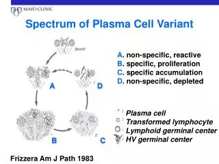
Plasma cells • Terminally differentiated cells of B-lymphocyte lineage • Produce antibodies • Normal plasma cells are incapable of dividing
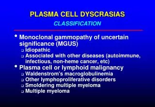
Classification of plasma cell disease • Multiple myeloma • Variants • Non-secretory myeloma • Indolant myeloma • Smoldering myeloma • Plasma cell leukemia
Classification of plasma cell diseases • Plasmocytoma • Solitary plasmocytoma • Multiple plasmocytoma • Primary amyloidosis • POEMS Syndrome • Waldenström’s Macroglobulinemia • Heavy Chain Diseases
Definition • B-cell malignancy characterised by abnormal proliferation of plasma cells able to produce a monoclonal immunoglobulin (M protein)
Incidence • 3-9 cases per 100,000 population/y • more frequent in elderly • modest male predominance
Clinical forms of MM • Multiple myeloma • Non-secretory myeloma • Smoldering myeloma • Plasma cell leukemia
M-protein (paraprotein) • Seen in 99% of cases in serum and/or urine • IgG > 50% • IgA 20-25% • IgE and IgD 1-3% • light chain 20% • 1% of cases are nonsecretory
Clinical manifestations are related to malignant behavior of plasma cells • and abnormalities produced by M protein
plasma cell proliferation • multiple osteolytic bone lesions • Hypercalcemia • bone marrow suppression ( pancytopenia ) • monoclonal M protein • decreased level of normal immunoglobulins • hyperviscosity
Symptoms • Bone pains • Weakness and fatigue • Weight loss
Laboratory • ESR > 100 • anaemia, thrombocytopenia • rouleaux in peripheral blood smears • marrow plasmacytosis • hyperproteinemia • hypercalcemia • proteinuria • azotemia
Causes of renal failure in MM • Hyperviscosity • Hypercalcemia • Hyperuricemia • Light chain deposition • Analgesic nephropathy
Evaluation for a suspected MM • Serum and urine protein electrophoresis • Serum and urine immunofixation and immunglobulin quantitation • Radiographic skeletal survey • Bone marrow examination
Diagnostic Criteria for Multiple Myeloma • Major criteria • I. Plasmacytoma on tissue biopsy • II. Bone marrow plasma cell > 30% • III. Monoclonal M spike on electrophoresis IgG > 3,5g/dl,IgA>2g/dl, light chain>1g/dl in 24h urinsample • Minor criteria • a. Bone marrow plasma cells 10-30% • b. M spike but less than above • c. Lytic bone lesions • d. Normal IgM < 50mg, IgA < 100mg, IgG < 600mg/dl Minimum of 1 major and 1 minor or 3 minor criteria including A and B
Staging of Multiple Myeloma Clinical staging • is based on level of haemoglobin, serum calcium, immunoglobulins and presence or not of lytic bone lesions • correlates with myeloma burden and prognosis I. Low tumor mass II. Intermediate tumor mass III. High tumor mass • subclassification A - creatinine < 2mg/dl B - creatinine > 2mg/dl
Poor prognosis factors • Cytogenetical abnormalities of 11 and 13 chromosomes • Beta-2 microglobulin > 2,5 ug/ml
Treatment • Patients <65-70 years • Velcade + Deksametazon • Thalidomid • VAD (Vincristin, Adriamycin, Dexamethasone) • high-dose therapy with autologous stem cell transplantation • allogeneic stem cell transplantation ( conventional and „mini”) • Patients >65 years • conventional chemotherapy
Treatment • Conventional chemotherapy • Velcade + dekasametazon • Talidomid • VAD (Vincristin, Adriamycin, Dexamethasone) • Melphlan + Prednisone • M2 ( Vincristine, Melphalan, Cyclophosphamid, BCNU, Prednisone) • Response rate 50-60% patients • Long term survival 5-10% patients
Treatment • Autologous transplantation • patients < 65-70 years • treatment related mortality 10-20% • response rate 80% • long term survival 40-50%
Treatment • non-myeloablative therapy and allogeneic transplantation
Treatment • Supportive treatment • biphosphonates, calcitonin • recombinant erythropoietin • immunoglobulins • plasma exchange • radiation therapy
Monoclonal gammopathy of undetermined significance ( MGUS) • M protein presence, stable • levels of M protein: IgG<3,5g IgA<2g,ligh chain<1g/day • normal immunoglobulins - normal levels • marrow plasmacytosis < 5% • complete blood count - normal • no lytic bone lesions • no signs of disease
Monoclonal gammopathy of undetermined significance ( MGUS) • M protein • 3% of people > 70 years • 15% of people > 90 years • 10% of patients with MGUS develop multiple myeloma
Diagnostic criteria for smoldering myeloma • Same as MGUS except: • Serum M-component at myeloma levels • Marrow plasmocytosis 10-30%
Plasma cell leukemia • >2x10⁹ plasma cells in peripheral blood • Younger age • Higher incidence of organomegaly and lymphadenopathy • More extensive bone marrow infiltration • Poor response to chemotherapy
Non-secretory myeloma • 1% of multiple myeloma • No serum or urine monoclonal protein • Must rule out IgD and IgE myeloma
Waldenström’s Macroglobulinemia • Monoclonal protein is IgM • No lytic lesions • Hyperviscosity (headache, tinnitus, dizziness, somnelence, etc) • Bone marrow aspiration reveals lymphoplasmocytic cells
Solitary plasmacytoma • Localized plasma cell tumor • Absence of plasma cell infiltrate in bone marrow biyopsy • No evidence of other lytic lesions on radiographic examination • Absence of renal failure, anemia or hypercalcemia
Osteosclerotic Myeloma(POEMS Syndrome) • Polyneuropathy • Organomegaly (hepatomegaly, LAP) • Endocrinopathy (hypogonadism, hypoyhtroidism) • Monoclonal gammopathy • Skin changes (hyperpigmentation)