Hematology
Hematology. Hematopoietic stem cells ( HSCs ) are multipotent stem cells that give rise to all the blood cell types including Myeloid ( monocytes and macrophages, neutrophils , basophils , eosinophils , erythrocytes, megakaryocytes /platelets, dendritic cells), and


Hematology
E N D
Presentation Transcript
Hematology Dr. Alka Stoelinga
Hematopoietic stem cells (HSCs) are multipotent stem cells that give rise to all the blood cell types including • Myeloid (monocytes and macrophages, neutrophils, basophils, eosinophils, erythrocytes, megakaryocytes/platelets, dendritic cells), and • Lymphoid lineages (T-cells, B-cells, NK-cells). • The hematopoietic tissue contains cells with long-term and short-term regeneration capacities and committed multipotent, oligopotent, and unipotent progenitors. • HSCs constitute 1:10.000 of cells in myeloid tissue. Dr. Alka Stoelinga
Source • HSCs are found in the bone marrow of adults, which includes femurs, hip, ribs, sternum, and other bones. • Cells can be obtained directly by removal from the hip bones using a needle and syringe, or from the blood following pre-treatment with cytokines, such as G-CSF (granulocyte colony-stimulating factors), that induce cells to be released from the bone marrow compartment. • Other sources for clinical and scientific use include umbilical cord blood, placenta, mobilized peripheral blood. Dr. Alka Stoelinga
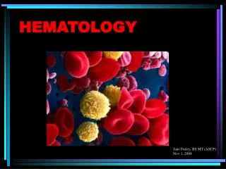
Erythrocytes • An erythrocyte has a disk diameter of 6–8 µm and a thickness of 2 µm, being much smaller than most other human cells. • These cells have a volume of about 90 fL with a surface of about 136 μm2, and can swell up to a sphere shape containing 150 fL, without membrane distension. • Adult humans have roughly 2–3 × 10¹³ (20-30 trillion) red blood cells at any given time, comprising approximately one quarter of the total human body cell number (women have about 4 to 5 million erythrocytes per microliter (cubic millimeter) of blood and men about 5 to 6 million • People living at high altitudes with low oxygen tension will have more. • Red blood cells are thus much more common than the other blood particles • There are about 4,000–11,000 white blood cells and about 150,000–400,000 platelets in each microliter of human blood. • Human red blood cells take on average 20 seconds to complete one cycle of circulation. Dr. Alka Stoelinga
Erythrocytes • As red blood cells contain no nucleus, protein biosynthesis is assumed to be absent in these cells, although a recent study indicates the presence of all the necessary biomachinery in human red blood cells for protein biosynthesis. • The blood’s red color is due to the spectral properties of the hemic iron ions in hemoglobin. • Each red blood cell contains approximately 270 million of these hemoglobin biomolecules, each carrying four heme groups • Hemoglobin comprises about a third of the total cell volume. • This protein is responsible for the transport of more than 98% of the oxygen (the remaining oxygen is carried dissolved in the blood plasma). • The red blood cells of an average adult male store collectively about 2.5 grams of iron, representing about 65% of the total iron contained in the body. Dr. Alka Stoelinga
Erythrocytes are produced by erythropoiesis, developing from committed stem cells to mature erythrocytes in about 7 days. • When matured, these cells live in blood circulation for about 100 to 120 days. • At the end of their lifespan, they become senescent, and are removed from circulation. Dr. Alka Stoelinga
Erythropoiesis • Erythropoiesis is the development process in which new erythrocytes are produced, through which each cell matures in about 7 days. • Through this process erythrocytes are continuously produced in the red bone marrow of large bones, at a rate of about 2 million per second in a healthy adult. • (In the embryo, the liver is the main site of red blood cell production.) • The production can be stimulated by the hormone erythropoietin (EPO), synthesized by the kidney. • Just before and after leaving the bone marrow, the developing cells are known as reticulocytes • These comprise about 1% of circulating red blood cells. Dr. Alka Stoelinga
Senescence • The aging erythrocyte undergoes changes in its plasma membrane, making it susceptible to selective recognition by macrophages and subsequent phagocytosis in the reticuloendothelial system (spleen, liver and bone marrow), thus removing old and defective cells and continually purging the blood. • This process is termed eryptosis, or erythrocyte programmed cell death. • This process normally occurs at the same rate of production by erythropoiesis, balancing the total circulating red blood cell count. • Eryptosis is increased in a wide variety of diseases including sepsis, haemolytic uremic syndrome, malaria, sickle cell anemia, beta-thalassemia, glucose-6-phosphate dehydrogenase deficiency, phosphate depletion, iron deficiency and Wilson's disease. • Breakdown products are recirculated in the body. • The heme constituent of hemoglobin are broken down into Fe3+ and biliverdin. • The biliverdin is reduced to bilirubin, which is released into the plasma and recirculated to the liver bound to albumin. • The iron is released into the plasma to be recirculated by a carrier protein called transferrin. • Almost all erythrocytes are removed in this manner from the circulation before they are old enough to hemolyze. • Hemolyzed hemoglobin is bound to a protein in plasma called haptoglobin Dr. Alka Stoelinga
Erythrocyte, Thrombocyte/ Platelet, Leukocyte RBC Dr. Alka Stoelinga
RBC abnormalities • Size • Anisocytosis • Macrocytosis • Microcytosis • Shape • Poikilocytosis • Spherocytosis • Elliptocytosis • Sickle cell • Staining • Hypochromia • Target cell Dr. Alka Stoelinga
RBC abnormalities • Size • Anisocytosis • Macrocytosis • Microcytosis Dr. Alka Stoelinga
Shape • Poikilocytosis • Spherocytosis • Elliptocytosis • Sickle cell Dr. Alka Stoelinga
RBC abnormalities • Staining • Hypochromia • Target cell Dr. Alka Stoelinga
3% and 5% Sodium Chloride Injection physiological saline or isotonic saline/ NS 0.9% 2%dextrose/ 0.45%Saline/ Dr. Alka Stoelinga
Normal Reference Ranges Dr. Alka Stoelinga
Normal Reference Ranges Dr. Alka Stoelinga
Normal Reference Ranges Dr. Alka Stoelinga
Major manifestations of hematological disorders • Anemia • Polycythaemia • Leucopenia • Leukocytosis • Lymphadenopathy • Splenomegaly • Bleeding • Thrombocytopenia • Thrombocytosis • Venous thrombosis • Abnormal coagulation screen • Pancytopenia • Infections Dr. Alka Stoelinga
ANEMIA • Anemia refers to a state in which the level of hemoglobin in blood is below the normal range appropriate for age and sex. Dr. Alka Stoelinga
Classification of Anemia • Etiological classification • Blood loss • Impaired production of RBC • Excessive destruction • Morphological classification • Macrocytic • Microcytic hypochromic • Normocytic normochromic Dr. Alka Stoelinga
Classification of Anemia • Morphological classification • Macrocytic MCV>94 MHCH>31 Megaloblasticdyspoiesis • Vit B12 deficiency- Pernicious anemia • Folic acid deficiency- Nutritional megaloblastic anemia, Sprue, Malabsorptions • Inborn errors- Oroticaciduria • Abnormal DNA synthesis- Chemotherapies, Anticonvulsants, OCP • Microcytic hypochromic MCV<80 MHCH<31 • Fe Deficincy- Chronic blood loss, Inadequate intake, Malabsorption, Increase demand • Abnormal globin synthesis- Thalassemia e/eouthemoglobinopathies • Abnormal Porphyrin and heme synthesis • Pyridoxine responsive anemia • Normocytic normochromic MCV 82-92 MCHC>30 • Blood loss • Increase plasma volume; Pregnancy, Overhydration • Hemolytic anemia • Hypoplastic marrow- Aplastic anemia, RBC aplasia • Infiltrate BM: Leukemia, MM, Myelofibrosis • Abnormal endocrine disorders: Hypothyroidism, Adrenal insufficiency • Liver disease, Kidney Disease, Cirrhosis Dr. Alka Stoelinga
Classification of Anemia • Etiological classification • Blood loss • Acute: Accidents/ GI bleed • Chronic:Hypermenorrhea/ Parasitic infections • Impaired production of RBC • Abnormal BM • Aplastic Anemia • Myelopthisis: Myelofibrosis, leukemia, • Essential factor deficiency • Deficiency anemia; Fe, Vit B12, Folic acid • Anemia in renal ds: Decreased erythropoietin • Stimulatory factor deficiency • A in chr. Ds • A. in hypopituitarism • A. in hypothyroidism • Excessive destruction • Intracorpuscular deficiency • Membrane: Heri. Spherocytosis/ ovalocytosis • Enzyme: G6PD / PK deficiency • Hb: Thalassemia, hemoglobinopathies • Extracorpuscular • Mechanical: March HA, Microangiopathic HA (MAHA) • Chemical/ Physical • Inf: Clostridium tetani • Antibodies: SLE • Hypersplenism Dr. Alka Stoelinga
Clinical features Dr. Alka Stoelinga
Clinical features Non- specific symptoms • Tiredness • Lightheadedness • Breathlessness • Dizziness/ Tinnitus/ Headache/ Dimness of vision • Ankle swelling • Worsening of any previous coexisting disease- Angina Non- Specific signs • Pallorness of Mucous membrane + Skin • Tachypnea/ Exertional dyspnea • Raised JVP • Flow murmurs • Ankle edema • Postural hypotension • Tachycardia Dr. Alka Stoelinga
Terminologies • Hb: is the iron-containing oxygen-transport metalloprotein in the red blood cells • HCT: The hematocrit (Ht or HCT) or packed cell volume (PCV) or erythrocyte volume fraction (EVF) is the proportion of blood volume that is occupied by red blood cells. • It is normally about 48% for men and 38% for women • MCV: The mean corpuscular volume, or "mean cell volume" (MCV), is a measure of the average red blood cell volume that is reported as part of a standard complete blood count (80-100 fL) • RDW: The red blood cell distribution width, RDW, or RCDW is a measure of the variation of red blood cell (RBC) width that is reported as part of a standard complete blood count. Usually red blood cells are a standard size of about 6-8μm • Retics:Retics are immature red blood cells, typically composing about 1% of the red cells in the human body Dr. Alka Stoelinga
INVESTIGATIONS Dr. Alka Stoelinga