GRAM NEGATIVE COCCI & UNCOMMON GRAM NEGATIVE BACILLI
750 likes | 1.82k Vues
GRAM NEGATIVE COCCI & UNCOMMON GRAM NEGATIVE BACILLI. Assoc. Prof.Dr.Yesim Gurol. Gram negative cocci and coccobacilli (2 hours): Learning Objectives. 1. Defines ‘’Gram negative cocci’’, ‘’Gram negative coccobacilli’’

GRAM NEGATIVE COCCI & UNCOMMON GRAM NEGATIVE BACILLI
E N D
Presentation Transcript
GRAM NEGATIVE COCCI& UNCOMMON GRAM NEGATIVE BACILLI Assoc. Prof.Dr.Yesim Gurol
Gram negative cocci and coccobacilli (2 hours):Learning Objectives 1. Defines ‘’Gram negative cocci’’, ‘’Gram negative coccobacilli’’ • 1.1 Lists Gram negative cocci and Gram negative coccobacilli in normal flora. • 1.2. Lists pathogenic Gram negative cocci and Gram negative coccobacilli for human. • 1.3. Lists virulance factors, defines tissue damage mechanisms. 2. Lists the clinical tables related with Gram negative cocci and Gram negative coccobacilli and defines pathogenetic mechanisms. • 2.1. Defines the clinical importance of Gram negative cocci and Gram negative coccobacilli (N.gonorrhoeae,N.meningitidis,Haemophilus spp, Aggregatibacter actinomycetemcomitans, Pasteurella multocida , Eikenella corrodens , Kingella kingae, Bordetella pertussis, Francisella tularensis,Brucella spp.) • 2.2. Lists the syndromes related with Neisseria spp. Like Waterhouse Friederichson, Fitz-Hugh-Curtis syndrome • 2.3. Lists the diagnostic methods of Gram negative cocci and Gram negative coccobacilli 2.4. Defines the prevention methods for infections related with Gram negative cocci and Gram negative coccobacilli
Neisseria meningitidis Biology, Virulence, and Disease • Gram-negative diplococci with fastidious growth requirements • Grows best at 35° C to 37° C in a humid atmosphere • Oxidase and catalase positive; acid produced from glucose and maltose oxidatively • Outer surface antigens include polysaccharide capsule, pili, and lipooligosaccharide (LOS) • Capsule protects bacteria from antibody-mediated phagocytosis • Specific receptors for meningococcal pili allow colonization of nasopharynx • Bacteria can survive intracellular killing in the absence of humoral immunity • Endotoxin mediates most clinical manifestations
Epidemiology • Humans are the only natural hosts • Person-to-person spread occurs via aerosolization of respiratory tract secretions • Highest incidence of disease is in children younger than 5 years, institutionalized people, and patients with late complement deficiencies • Meningitis and meningococcemia most commonly caused by serogroups B, C, and Y; pneumonia most commonly caused by serogroups Y and W135; serogroups A and W135 associated with disease in underdeveloped countries • Disease occurs worldwide, most commonly in the dry, cold months of the year
Diagnosis • Gram stain of cerebrospinal fluid is sensitive and specific but is of limited value for blood specimens (too few organisms are generally present, except in overwhelming sepsis) • Culture is definitive, but organism is fastidious and dies rapidly when exposed to cold or dry conditions • Tests to detect meningococcal antigens are insensitive and nonspecific Treatment, Prevention, and Control • Breast-fed infants have passive immunity (first 6 months) • Treatment is with penicillin (drug of choice) • Chemoprophylaxis for contact with persons with the disease is with rifampin, ciprofloxacin, or ceftriaxone • For immunoprophylaxis, vaccination is an adjunct to chemoprophylaxis; it is used only for serogroups A, C, Y, and W135; no effective vaccine is available for serogroup B
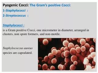
Neisseria species are aerobic, Gram-negative bacteria, typically coccoid shaped (0.6 to 1.0 μm in diameter) and arranged in pairs (diplococci) with adjacent sides flattened together (resembling coffee beans). • The bacteria are not motile and do not form endospores. • All species are oxidase positive, and most produce catalase-properties that combined with the Gram-stain morphology allow for a rapid, presumptive identification of a clinical isolate. Acid is produced by oxidation of carbohydrates (not by fermentation). • N. gonorrhoeae strains produce acid by oxidizing glucose, and N. meningitidis strains oxidize both glucose and maltose. Other carbohydrates are not oxidized. This pattern of carbohydrate utilization is useful for differentiating these pathogenic strains from other Neisseria species.
Nonpathogenic species of Neisseria can grow on nutrient agar incubated at 35° C to 37° C. In contrast, N. meningitidis has variable growth on nutrient agar, and N. gonorrhoeae is a fastidious organism, requiring complex media for growth; it is adversely affected by exposure to dry conditions or fatty acids. • All strains of N. gonorrhoeae require cystine and an energy source (e.g., glucose, pyruvate, and lactate) for growth, and many strains require supplementation of media with amino acids, purines, pyrimidines, and vitamins. Soluble starch is added to the media to neutralize the toxic effect of the fatty acids. • Thus N. gonorrhoeae does not grow on blood agar but does grow on chocolate agar and other enriched supplemented media. The optimum growth temperature is 35° C to 37° C, with poor survival of the organism at cooler temperatures. • A humid atmosphere supplemented with 5% carbon dioxide (CO2) is either required or enhances growth of N. gonorrhoeae. Although the fastidious nature of this organism makes recovery from clinical specimens difficult, it is nevertheless easy for the organism to be transmitted sexually from person to person.
The structure of N. gonorrhoeae and N. meningitidis is typical of gram-negative bacteria, with the thin peptidoglycan layer sandwiched between the inner cytoplasmic membrane and the outer membrane. • The major virulence factor for N. meningitidis is the polysaccharide capsule. • Although the outer surface of N. gonorrhoeae is not covered with a true carbohydrate capsule, the cell surface of N. gonorrhoeae has a capsule-like negative charge. • Antigenic differences in the polysaccharide capsule of N. meningitidis are the basis for serogrouping these bacteria. Thirteen serogroups are currently recognized (A, B, C, D, H, I, K, L, W-135, X, Y, Z, 29E), with most infections caused by serogroups A, B, C, Y, and W135.
Pathogenic and nonpathogenic strains of Neisseria have pili that extend from the cytoplasmic membrane through the outer membrane. Pili mediate a number of functions, including attachment to host cells, transfer of genetic material, and motility, and the presence of pili in N. gonorrhoeae and N. meningitidis appears to be important for pathogenesis. • The pili are composed of repeating protein subunits (pilins), whose expression is controlled by the pil gene complex. Pili expression is associated with virulence, in part because the pili mediate attachment to nonciliated epithelial cells and provide resistance to killing by neutrophils. • Pilin proteins have a conserved region at the amino terminal end and a highly variable region at the exposed carboxyl terminus. The carboxyl terminal portion of the pilin protein can be phosphorylated and glycosylated and is associated with a second protein, PilC, which contributes to its antigenic diversity. • The lack of immunity to reinfection with N. gonorrhoeae results partially from the antigenic variation among the pilin proteins and partially from the phase variation in pilin expression; these factors complicate attempts to develop effective vaccines for gonorrhea.
Gonorrhea occurs naturally only in humans; it has no other known reservoir. • It is second only to chlamydia as the most commonly reported sexually transmitted disease in the United States. Infection rates are the same in males and females, are disproportionately higher in blacks than in Hispanic Americans and whites, and are highest in the southeastern United States. • The peak incidence of the disease is in the age group 15 to 24 years. The incidence of disease decreased from 1978 to 1997; however, between 1998 and 2006, the incidence of gonorrhea has remained relatively constant. • In 2006, almost 360,000 new infections were reported in the United States. • However, even this large number is an underestimation of the true incidence of disease because the diagnosis and reporting of gonococcal infections are incomplete. • Public health officials believe that at least half of the new infections are not reported.
N. gonorrhoeae is transmitted primarily by sexual contact. Women have a 50% risk of acquiring the infection as the result of a single exposure to an infected man, whereas men have a risk of approximately 20% as the result of a single exposure to an infected woman. • The risk of infection rises as the person has more sexual encounters with infected partners. • The major reservoir for gonococci is the asymptomatically infected person. Asymptomatic carriage is more common in women than in men.
As many as half of all infected women have mild or asymptomatic infections, whereas most men are initially symptomatic. • The symptoms generally clear within a few weeks in people with untreated disease, and asymptomatic carriage may then become established. • The site of infection also determines whether carriage occurs, with rectal and pharyngeal infections more commonly asymptomatic than genital infections.
Endemic meningococcal disease occurs worldwide, and epidemics are common in developing countries. Epidemic spread of disease results from the introduction of a new, virulent strain into an immunologically naïve population. Pandemics of disease have been uncommon in developed countries since World War II. • Of the 13 serogroups, almost all infections are caused by serogroups A, B, C, Y, and W135. In Europe and the Americas, serogroups B, C, and Y predominate in meningitis or meningococcemia; serogroups A and W135 predominate in developing countries. • Serogroups Y and W135 are most commonly associated with meningococcal pneumonia. N. meningitidis is transmitted by respiratory droplets among people in prolonged close contact, such as family members living in the same household and soldiers living together in military barracks.
Classmates in schools and hospital employees are not considered close contacts and are not at significantly higher risk of acquiring the disease, unless they are in direct contact with the respiratory secretions of an infected person. • Humans are the only natural carriers for N. meningitidis. Studies of the asymptomatic carriage of N. meningitidis have shown that there is a tremendous variation in its prevalence, from less than 1% to almost 40%. • The oral and nasopharyngeal carriage rates are highest for school-age children and young adults, are higher in lower socioeconomic populations (caused by person-to-person spread in crowded areas), and do not vary with the seasons, even though disease is most common during the dry, cold months of the year. • Carriage is typically transient, with clearance occurring after specific antibodies develop. Endemic disease is most common in children younger than 5 years, particularly infants, and teenagers and young adults. • People who are immunocompromised, the elderly, or those who live in closed populations (e.g., military barracks, prisons) are prone to infection during epidemics.
Neisseria gonorrhoeae • Gonorrhea: characterized by purulent discharge for involved site (e.g., urethra, cervix, epididymis, prostate, anus) after 2- to 5-day incubation period • Disseminated infections: spread of infection from genitourinary tract through blood to skin or joints; characterized by pustular rash with erythematous base and suppurative arthritis in involved joints • Ophthalmia neonatorum: purulent ocular infection acquired by neonate at birth Neisseria meningitidis • Meningitis: purulent inflammation of meninges associated with headache, meningeal signs, and fever; high mortality rate unless promptly treated with effective antibiotics • Meningococcemia: disseminated infection characterized by thrombosis of small blood vessels and multiorgan involvement; small, petechial skin lesions coalesce into larger hemorrhagic lesions • Pneumonia: milder form of meningococcal disease characterized by bronchopneumonia in patients with underlying pulmonary disease Eikenella corrodens • Human bite wounds: infection associated with traumatic (e.g., bite, fistfight injury) introduction of oral organisms into deep tissue • Subacute endocarditis: infection of endocardium characterized by gradual onset of low grade fevers, night sweats, and chills Kingella kingae • Subacute endocarditis: as with E. corrodens
Meningococcemia • Septicemia (meningococcemia) with or without meningitis is a life-threatening disease. • Thrombosis of small blood vessels and multiorgan involvement are the characteristic clinical features. • Small, petechial skin lesions on the trunk and lower extremities are common and may coalesce to form larger hemorrhagic lesions . • Overwhelming disseminated intravascular coagulation with shock, together with the bilateral destruction of the adrenal glands (Waterhouse-Friderichsen syndrome), may ensue. • A milder, chronic septicemia has also been observed. • Bacteremia can persist for days or weeks, and the only signs of infection are a low-grade fever, arthritis, and petechial skin lesions. • The response to antibiotic therapy in patients with this form of the disease is generally excellent.
Additional infections caused by N. meningitidis • pneumonia, • arthritis, • urethritis. • Meningococcal pneumonia is usually preceded by a respiratory tract infection. Symptoms include cough, chest pain, rales, fever, and chills. Evidence of pharyngitis is observed in most affected patients. The prognosis in patients with meningococcal pneumonia is good. Other diseases associated with N. gonorrhoeae are perihepatitis (Fitz-Hugh-Curtis syndrome); purulent conjunctivitis , particularly in newborns infected during vaginal delivery (ophthalmia neonatorum); anorectal gonorrhea in homosexual men; and pharyngitis.
Microscopy • Gram stain is very sensitive (greater than 90%) and specific (98%) in detecting gonococcal infection in men with purulent urethritis . • Its sensitivity in detecting infection in asymptomatic men is 60% or less. • The test is also relatively insensitive in detecting gonococcal cervicitis in both symptomatic and asymptomatic women, although a positive result is considered reliable when an experienced microscopist sees gram-negative diplococci within polymorphonuclear leukocytes. • Gram stain can be reliably used to diagnose infections in men with purulent urethritis and women with cervicitis, but all negative results in women and asymptomatic men must be confirmed by culture. • Gram stain is also useful for the early diagnosis of purulent arthritis but is insensitive and nonspecific for the detection of N. gonorrhoeae in patients with skin lesions, anorectal infections, or pharyngitis. • Commensal Neisseria species in the oropharynx and morphologically similar bacteria in the gastrointestinal tract can be confused with N. gonorrhoeae. • N. meningitidis can be readily seen in the cerebrospinal fluid (CSF) of patients with meningitis unless the patient has received prior antimicrobial therapy. • Most patients with bacteremia caused by other organisms have so few organisms present in their blood that the Gram stain has no value. • In contrast, patients with overwhelming meningococcal disease commonly have large numbers of organisms in their blood, which can be seen when the peripheral blood leukocytes are Gram stained.
Antigen testing for the detection of N. Gonorrhoeae • is less sensitive than culture or nucleic acid amplification tests • not recommended unless confirmatory tests are performed on negative specimens. • Commercial tests to detect N. meningitidis capsular antigens in CSF, blood, and urine (where the antigens are excreted) were widely used in the past but have fallen into disfavor in recent years because the tests are less sensitive than Gram stains, and false-positive reactions, particularly with urine specimens, can occur. Nucleic acid amplification (NAA) assays specific for N. gonorrhoeae have been developed for the direct detection of bacteria in clinical specimens. Tests using these assays are sensitive, specific, and rapid (results are available in 4 hours). Combination NAA assays for both N. gonorrhoeae and Chlamydia organisms are available and have replaced culture in most labs. The primary problem with this approach is that it cannot be used to monitor antibiotic resistance of the identified pathogens.
Culture N. gonorrhoeae can be readily isolated from genital specimens if care is taken in collecting and processing the specimens .Because other commensal organisms normally colonize mucosal surfaces, all genital, rectal, and pharyngeal specimens must be inoculated onto both non-selective media (e.g., chocolate blood agar) and selective media that suppress the growth of contaminating organisms (e.g., modified Thayer-Martin medium). A nonselective medium should be used because some gonococcal strains are inhibited by the vancomycin present in most selective media. The gonococci die rapidly if specimens are allowed to dry. Therefore drying and cold temperatures should be avoided by directly inoculating the specimen onto prewarmed media at the time of collection.
The endocervix must be properly exposed to ensure that an adequate specimen is collected. Although the endocervix is the most common site of infection in women, the rectal specimen may be the only one positive for gonococci in women who have asymptomatic infections, as well as in homosexual and bisexual men. Blood culture results are generally positive for gonococci only during the first week of the infection in patients with disseminated disease. In addition, special handling of blood specimens is required to ensure the adequate recovery of gonococci, because supplements present in the blood culture media can be toxic to Neisseria. Culture results of specimens from infected joints are positive for the organism if the specimens are collected at the time the arthritis develops, but skin specimen cultures are generally unrewarding.
Vaccines • directed against the group-specific capsular polysaccharides have been developed for antibody-mediated immunoprophylaxis. • A polyvalent polysaccharide-protein conjugate vaccine effective against serogroups A, C, Y, and W135 was licensed in the United States in 2005. In 2007, the Advisory Committee on Immunization Practices (ACIP) recommended routine vaccination with one dose of this vaccine for all persons aged 11 to 18 years and other persons at increased risk for meningococcal disease. • Unfortunately the group B polysaccharide is a weak immunogen and cannot induce a protective antibody response. • Thus immunity to group B N. meningitidis must develop naturally after exposure to cross-reacting antigens. • Vaccination with a suspension containing serogroup A can be used for control of an outbreak of disease, for travelers to hyperendemic areas, and for people at increased risk for disease (e.g., patients with complement deficiency).
Eikenella corrodens • In the early 1960s, a collection of small, fastidious, gram-negative rods were classified by workers at the CDC as members of the HB group (named after the patient infected with the original isolate). • The organisms were subsequently subdivided into subgroup HB-1 (now known as Eikenella corrodens), subgroup HB-2 (Aggregatibacter [Haemophilus] aphrophilus), and subgroups HB-3 and HB-4 (Aggregatibacter [Actinobacillus] actinomycetemcomitans). • In addition to being morphologically similar, these organisms colonize the human oropharynx; in the setting of preexisting heart disease, they can cause subacute bacterial endocarditis. In fact, the group of fastidious, gram-negative rods associated with subacute endocarditis is known by the now taxonomically incorrect acronym HACEK (H. aphrophilus, A. actinomycetemcomitans, Cardiobacterium hominis, E. corrodens, and K. kingae).
E. corrodens is a moderate-sized (0.2 × 2.0 μm), nonmotile, non-spore-forming, facultatively anaerobic, gram-negative rod. The organism is named after Eiken, who characterized the bacterium and observed the ability of the organism to pit or "corrode" agar (from its ability to split polygalacturonic acid). E. corrodens is a normal inhabitant of the human upper respiratory tract, but because of its fastidious growth requirements, it is difficult to detect unless specific selective culture media are used.
It is an opportunistic pathogen that causes infections in patients who are immunocompromised or have diseases or trauma of the oral cavity. • E. corrodens is most commonly isolated in the settings of a human bite wound or fistfight injury. Other infections • endocarditis, • sinusitis, • meningitis, • brain abscesses, • pneumonia, • lung abscesses.
Because most infections originate from the oropharynx, polymicrobial mixtures of aerobic and anaerobic bacteria are often present in cultures. • A slow-growing, fastidious organism, E. corrodens requires 5% to 10% carbon dioxide to grow. Small (0.5 to 1 mm) colonies are observed after 48 hours of incubation on blood or chocolate agar, but the organism grows poorly or not at all on selective media for gram-negative rods. • Pitting in agar is a useful differential characteristic, but fewer than half of all isolates exhibit pitting. The organism also produces a characteristic bleachlike odor. Thus if a slow-growing, gram-negative rod is found to pit blood agar and produce a bleachlike order, a preliminary identification of the organism can be made. • E. corrodens is resistant to many antibiotics that are selected empirically to treat bite-wound infections.
Kingella kingae • Kingella species are small, gram-negative coccobacilli that morphologically resemble Neisseria species and reside in the human oropharynx. The bacteria are facultatively anaerobic, ferment carbohydrates, and have fastidious growth requirements. • K. kingae, the most commonly isolated species, has been primarily responsible for septic arthritis in children and endocarditis in patients of all ages. Because the organism grows slowly, it may take 3 or more days of incubation for the organism to be detected in clinical specimens. • Most strains are susceptible to β-lactam antibiotics, including penicillin, tetracyclines, erythromycin, fluoroquinolones, and aminoglycosides.
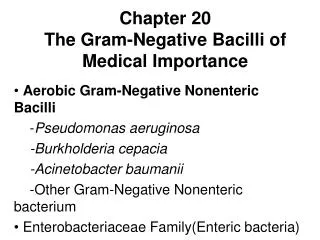
Pasteurellaceae Haemophilus influenzae • Meningitis: primarily a disease of unimmunized children; characterized by fever, severe headache, and systemic signs • Epiglottitis: primarily a disease of unimmunized children; characterized by initial pharyngitis, fever, and difficulty breathing, and progressing to cellulitis and swelling of the supraglottic tissues, with obstruction of the airways possible • Pneumonia: inflammation and consolidation of the lungs observed primarily in the elderly with underlying chronic pulmonary disease; typically caused by nontypeable strains Haemophilus aegyptius • Conjunctivitis: an acute, purulent conjunctivitis ("pink eye") Haemophilus ducreyi • Chancroid: sexually transmitted disease characterized by a tender papule with an erythematous base, progressing to painful ulceration with associated lymphadenopathy Aggregatibacter actinomycetemcomitans • Endocarditis: responsible for subacute form of endocarditis in patients with underlying damage to the heart valve Aggregatibacter aphrophilus • Endocarditis: as with A. actinomycetemcomitans Pasteurella multocida • Bite wound: most common manifestation is infected cat- or dog-bite wound; particularly common with cat bites, because the wounds are deep and difficult to disinfect
Haemophilae are small, sometimes pleomorphic, gram-negative rods present on the mucous membranes of humans. • Haemophilus influenzae is the species most commonly associated with disease, with infections most often reported in pediatric patients before the introduction of the H. influenzae type b (HIB) vaccine. • Haemophilus aegyptius is an important cause of acute, purulent conjunctivitis. • Haemophilus ducreyi is well recognized as the etiologic agent of the sexually transmitted disease soft chancre, or chancroid. • The other members of the genus are commonly isolated in clinical specimens (e.g., Haemophilus parainfluenzae is the most common species in the mouth) but are rarely pathogenic, being responsible primarily for opportunistic infections.
The growth of most species of Haemophilus requires supplementation of media with one or both of the following growth-stimulating factors: • hemin (also called X factor for unknown factor), • nicotinamide adenine dinucleotide (NAD; also called V factor for "vitamin"). • Although both factors are present in blood-enriched media, sheep blood agar must be gently heated to destroy the inhibitors of V factor. For this reason, heated blood ("chocolate") agar is used for the in vitro isolation of Haemophilus.
Lipopolysaccharide with endotoxin activity is present in the cell wall, and strain-specific and species-specific proteins are found in the outer membrane. • The surface of many but not all strains of H. influenzae is covered with a polysaccharide capsule, and six antigenic serotypes (a through f) have been identified. • Before the introduction of the HIB vaccine, H. influenzae serotype b was responsible for more than 95% of all invasive Haemophilus infections. After the introduction of the vaccine, most disease caused by this serotype disappeared, and more than half of all invasive disease is now caused by nonencapsulated (nontypeable) strains.
Haemophilus Biology, Virulence, and Disease • Small, pleomorphic, gram-negative rods or coccobacilli • Facultative anaerobes, fermentative • Most species require X and/or V factor for growth • H. influenzae subdivided serologically (types a to f) and biochemically (biotypes I to VIII) • H. influenzae type b is clinically most virulent (with PRP, [polyribitol phosphate] in capsule) • Haemophilus adhere to host cells via pili and nonpilus structures Epidemiology • Haemophilus species commonly colonized in humans although encapsulated Haemophilus species, particularly H. influenzae type b, are uncommon members of normal flora • Disease caused by H. influenzae type b was primarily a pediatric problem; eliminated in immunized populations • H. ducreyi disease is uncommon in the United States • With the exception of H. ducreyi, which is spread by sexual contact, most Haemophilus infections are caused by the patient's oropharyngeal flora (endogenous infections) • Patients at greatest risk for disease are those with inadequate levels of protective antibodies, those with depleted complement, and those who have undergone splenectomy
Diagnosis • Microscopy is a sensitive test for detecting H. influenzae in cerebrospinal fluid (CSF), synovial fluid, and lower respiratory specimens but not from other sites • Culture is performed using chocolate agar • Antigen tests are specific for H. influenzae type b; therefore, these tests are nonreactive for infections caused by other organisms Treatment, Prevention, and Control • Haemophilus infections are treated with broad-spectrum cephalosporins, azithromycin, or fluoroquinolones; many strains are resistant to ampicillin • Active immunization with conjugated PRP vaccines prevents most H. influenzae type b infections
Laboratory Diagnosis • Specimen Collection and Transport • Because most Haemophilus infections in vaccinated individuals originate from the oropharynx and are restricted to the upper and lower respiratory tract, contamination of the specimen with oral secretions should be avoided. • Direct needle aspiration should be used for the microbiologic diagnosis of sinusitis or otitis, and sputum produced from the lower airways is used for the diagnosis of pneumonia. • Culture of blood for patients with pneumonia may be useful but would be predictably negative in patients with upper respiratory infections. • Both blood and cerebrospinal fluid (CSF) should be collected from nonimmune children with the diagnosis of meningitis. Because there are approximately 107 bacteria per mL of CSF in patients with untreated meningitis, 1 to 2 mL of fluid is generally adequate for microscopy, culture, and antigen-detection tests. • Microscopy and culture are less sensitive if the patient has been exposed to antibiotics before the CSF is collected.
Blood cultures should also be collected for the diagnosis of epiglottitis, cellulitis, and arthritis. Specimens should not be collected from the posterior pharynx in patients with suspected epiglottitis because the procedure may stimulate coughing and obstruct the airway. • Specimens for the detection of H. ducreyi should be collected with a moistened swab from the base or margin of the ulcer. Culture of pus collected by aspiration from an enlarged lymph node can be performed but is generally less sensitive than culture of the ulcer. • The laboratory should be notified that H. ducreyi is suspected, because special culture techniques must be used for recovery of the organism.
If microscopy is performed carefully, the detection of Haemophilus species in clinical specimens is both sensitive and specific. Gram-negative rods ranging in shape from coccobacilli to long, pleomorphic filaments can be detected in more than 80% of CSF specimens from patients with untreated Haemophilus meningitis. • The microscopic examination of Gram-stained specimens is also useful for the rapid diagnosis of the organism in arthritis and lower respiratory tract disease. • Chocolate agar or Levinthal agar is used in most laboratories. However, if chocolate agar is overheated during preparation, V factor is destroyed, and Haemophilus species requiring this growth factor (e.g., H. influenzae, H. aegyptius, H. parainfluenzae) will not grow. • The bacteria appear as 1- to 2-mm, smooth, opaque colonies after 24 hours of incubation. They can also be detected growing around colonies of Staphylococcus aureus on unheated blood agar (satellite phenomenon). • The staphylococci provide the requisite growth factors by lysing the erythrocytes in the medium and releasing intracellular heme (X factor) and excreting NAD (V factor). The colonies of H. influenzae in these cultures are much smaller than they are on chocolate agar, because the V factor inhibitors present in blood are not inactivated.
Actinobacillus species are small, facultatively anaerobic, gram-negative rods that grow slowly (generally requiring 2 to 3 days of incubation). Actinobacillus actinomycetemcomitans was the most important human pathogen in this genus; however, in 2006 this species and Haemophilus aphrophilus were transferred into a new genus, Aggregatibacter. The remaining members of the genus Actinobacillus colonize the oropharynx of humans and animals and are rare causes of periodontitis, endocarditis, bite wound infections, and opportunistic infections. Two members of this genus are important human pathogens: • A. actinomycetemcomitans and A. aphrophilus Both species colonize the human mouth and can spread from the mouth into the blood and then stick to a previously damaged heart valve or artificial valve, leading to the development of endocarditis. • Endocarditis caused by these bacteria is particularly difficult to diagnose, because clinical signs and symptoms develop slowly (subacute endocarditis), and the bacteria grow slowly in blood cultures. Both species form adherent colonies that can be observed on the glass surface of the blood culture bottles and on agar media. The treatment of choice for endocarditis caused by these organisms is a cephalosporin such as ceftriaxone.
Pasteurella are small, facultatively anaerobic, fermentative coccobacilli commonly found as commensals in the oropharynx of healthy animals. • Most human infections result from animal contact (e.g., animal bites, scratches, shared food). • Pasteurella multocida (the most common isolate) and Pasteurella canis are human pathogens; the other Pasteurella species are rarely associated with human infections. • The following three general forms of disease are reported: • (1) localized cellulitis and lymphadenitis that occur after an animal bite or scratch (P. multocida from contact with cats or dogs; P. canis from dogs); • (2) an exacerbation of chronic respiratory disease in patients with underlying pulmonary dysfunction (presumably related to colonization of the patient's oropharynx followed by the aspiration of oral secretions); • (3) a systemic infection in immunocompromised patients, particularly those with underlying hepatic disease.
Bordetella is an extremely small (0.2 to 0.5 × 1 μm), strictly aerobic, gram-negative coccobacillus. Eight species are currently recognized, with four species responsible for human disease Bordetella pertussis ,the agent responsible for pertussis or whooping cough; Bordetella parapertussis, responsible for a milder form of pertussis; Bordetella bronchiseptica, responsible for respiratory disease in dogs, swine, laboratory animals, and occasionally respiratory disease in humans; and Bordetella holmesii, an uncommon cause of sepsis. Bordetella pertussis • After a 7- to 10-day incubation period, disease is characterized by the catarrhal stage (resembles the common cold), progressing to the paroxysmal stage (repetitive coughs followed by inspiratory whoops), then the convalescence stage (diminishing paroxysms and secondary complications) Bordetella parapertussis • Produces a milder form of pertussis Bordetella bronchiseptica • Primarily a respiratory disease of animals but can cause bronchopneumonia in humans Bordetella holmesii • Uncommon cause of sepsis
A direct fluorescent antibody procedure using either monoclonal or polyclonal antibodies can be used to examine specimens. • In this method, the aspirated specimen is smeared onto a microscopic slide, air-dried and heat fixed, and then stained with fluorescein-labeled antibodies directed against B. pertussis. • Antibodies against B. parapertussis should also be used to detect mild forms of pertussis caused by this organism. The direct fluorescent antibody test results are positive in about half of the patients with pertussis, but false-positive results can occur as a result of cross-reactions with other bacteria. Because of the sensitivity and specificity problems with this test, PCR and/or culture should also be performed. • Nucleic acid amplification methods such as polymerase chain reaction (PCR) are the most sensitive diagnostic test available for pertussis.
These methods have replaced microscopy and culture in most laboratories that offer laboratory testing. • At the present time, culture is generally offered by laboratories unable to perform nucleic-acid-based assays or in conjunction with these assays. The sensitivity of culture is affected by patient factors (i.e., stage of illness, use of antibiotics), the quality of the specimen, transport conditions, and culture methods. • The traditional use of Bordet-Gengou medium has been replaced by Regan-Lowe charcoal medium supplemented with glycerol, peptones, and horse blood. The media should be incubated in air at 35° C and in a humidified chamber. • Prolonged incubation (e.g., 7 to 12 days) is necessary. Because the quality of the media dramatically affects the success of culture, laboratories that infrequently culture specimens for Bordetella should arrange for the state public health department to process these specimens. Despite use of optimized culture conditions, fewer than half the infected patients have positive cultures
Francisella and Brucella are important zoonotic pathogens that occasionally cause human disease . • These organisms have also gained notoriety as potential agents of bioterrorism. Although the organisms have some common properties (e.g., very small coccobacilli, fastidious and slow growth requirements, always pathogenic in humans), they are taxonomically unrelated.
The genus Francisella consists of two species, Francisella tularensis and Francisella philomiragia. F. tularensis is the causative agent of tularemia (also called glandular fever, rabbit fever, tick fever, and deer fly fever) in animals and humans. F. tularensis is subdivided into four subspecies that are subdivided based on their biochemical properties. F. tularensis is a very small (0.2 × 0.2 to 0.7 μm), faintly staining, gram-negative coccobacillus. The organism is nonmotile, has a thin lipid capsule, and has fastidious growth requirements (i.e., most strains require cysteine for growth). It is strictly aerobic and requires 3 or more days before growth is detected in culture.
Francisella tularensis Biology, Virulence, and Disease • Very small, gram-negative coccobacilli (0.2 × 0.2 to 0.7 μm) • Strict aerobe; nonfermenter • Requires specialized media and prolonged incubation for growth in culture • Antiphagocytic capsule • Intracellular pathogen resistant to killing in serum and by phagocytes • Clinical symptoms and prognosis determined by route of infection: ulceroglandular, oculoglandular, glandular, typhoidal, oropharyngeal, gastrointestinal, pneumonic Epidemiology • Wild mammals, domestic animals, birds, fish, and blood-sucking arthropods are reservoirs; rabbits, cats, hard ticks, and biting flies are most commonly associated with human disease; humans are accidental hosts • Worldwide distribution; most common in United States in Oklahoma, Missouri, and Arkansas • Approximately 100 cases seen in United States, although the actual number may be much higher • The infectious dose is small when exposure is by arthropod, through skin, or by inhalation; large numbers of organisms must be ingested for infection by this route