Download
1 / 51
510 likes | 657 Vues
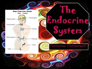
This chapter provides an overview of the endocrine system, its glands, and the hormones they produce. It discusses the differences between endocrine and exocrine glands, explaining how hormones are secreted into circulation and their slow response in target cells. The text covers various glands including the pituitary, thyroid, and adrenal glands, detailing the roles of hormones in regulating growth, metabolism, and homeostasis. Additionally, it explains hormone chemistry, mechanisms of action, and regulation of secretion, emphasizing the critical connection between the nervous and endocrine systems.

E N D
The Endocrine System Chapter 13
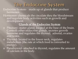
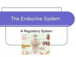
Endocrine • to interstitial fluid circulation • exocrine- secreted to ducts lumen or outside the body • Endocrine glands: • Pituitary, thyroid, parathyroid, adrenal & pineal • Hormone secretion + other functions: • Hypothalamus, thymus, pancreas, ovaries, testes, kidneys, stomach, liver, small intestine, skin, heart, adipose tissue & placenta
Hormone Operation • General chemical signal in circulation • Slower than nerve responses • Target cells must have a specific receptor • Response determined by responding cell, i.e. different cells may respond differently to the same hormone • Cell may respond to more than one hormone, • i.e. has more than one type receptor
Hormone Chemistry • Soluble in lipids = Hydrophobic • steroids, e.g. testosterone, estrogens, etc. • thyroid hormones, e.g. T3, T4 • Nitric oxide (NO) • Water soluble= Hydrophillic • Amino acid derivatives, e.g. epinephrine, norepinephrine • Peptides, e.g. antidiuretic Hormone (ADH), oxytocin • Proteins, e.g. insulin & growth hormone • General Action depends on chemistry
Lipid Soluble Action • Hormone detaches from carrier in blood stream • Diffusion through interstitial fluid & cell membrane into cell • Binds to & activates receptor • Receptor-hormone complex alters gene expression • If new mRNA protein synthesis • New proteins alter cell activity
Water-Soluble Action • Diffuses from blood and binds to receptor in plasma membrane • Starts reaction inside cell forming second messenger • Cyclic AMP is a common one • Second messenger causes activation of several proteins (enzymes) • Activated proteins produce physiological responses • Second messenger is inactivated
Control of Secretions • Release occurs in short bursts • Regulated by: • Signals from nervous system, e.g. adrenal medulla release of epinephrine • Chemical changes in blood, e.g Blood Ca2+ affects parathyroid hormone • Other hormones, e.g. ACTH from pituitary stimulates cortisol release from adrenal cortex
Hypothalamus & Pituitary • Major link between nervous & endocrine systems • Hypothalamic Cells synthesize at least 9 hormones • Pituitary synthesizes 7 • Regulate growth, development, metabolism & homeostasis
Pituitary • Two lobes; anterior & posterior • Hypophyseal portal veins • Connect capillaries in hypothalamus to capillaries in anterior pituitary
Hypothalamus Pituitary • Axons of hypothalamic neurons (neurosecretory cells) end near capillaries of hypothalamus • Secrete Releasing hormones or Inhibiting hormones portal veins • Regulate release of anterior pituitary hormones
Human Growth Hormone (hGH) • Promotes synthesis of IGFs = somatomedins • in liver, muscle, cartilage & bone • Released in bursts (~2 hour intervals) • Hypothalamus Growth Hormone Releasing Hormone (GHRH) & Growth Hormone Inhibiting Hormone (GHIH ) • Regulated by blood glucose levels
Thyroid Stimulating Hormone • Stimulates the formation & secretion of Thyroid hormones from thyroid gland • Hypothalamus Thyrotropin Releasing Hormone (TRH)- no TIH • Regulated by circulating thyroid hormone levels
Follicle Stimulating Hormone (FSH) & Luteinizing Hormone (LH) • In females: • FSH starts follicle development • LH stimulates formation of corpus luteum & secretion of progesterone • In males: • FSH stimulates sperm production in testes • LH stimulates release of testosterone • Gonadotrophin releasing Hormone (GnRH) from hypothalamus is suppressed by high levels of estrogen in females and testosterone in males
Prolactin (PRL) • Initiates & maintains milk production by mammary glands • Ejection of milk depends on oxytocin • Prolactin inhibiting hormone (PIH) suppresses prolactin release • High levels of Estrogens PRH prolactin release • Unknown function in males • Hypersecretion impotence
Adrenocortcotrophic Hormone (ACTH) • Controls production & secretion of glucocorticoids from adrenal cortex • Corticotrophin Releasing Hormone (CRH) from hypothalamus stimulates secretion of ACTH • Stress related stimuli can also stimulate ACTH release • Glucocorticoids inhibit CRH & ACTH release
Melanocyte Stimulating Hormone (MSH) • Small circulating amounts • Excess causes skin darkening
Posterior Pituitary • axon terminals from hypothalamus- • Release hormones • Oxytocin- enhance smooth muscle contraction during birth & milk ejection • may play role in emotional bonding • Antidiuretic Hormone (ADH) = vasopressin • Causes kidney to retain more water • Vasoconstriction increase in blood pressure • high blood osmotic pressure increase secretion
Thyroid Gland • Below larynx- two lobes • follicular cells surround follicles • thyroxin (T4) & triiodothyronine (T3) • Stored in follicle • Parafollicular cells (C-cells) • calcitonin
Thyroid Hormones • T4 & T3 increase basal metabolic rate, protein synthesis & growth • Blood level is controlled via feedback through hypothalamus • Increased body ATP demand can also raise blood levels • Calcitonin inhibits osteoclasts decrease in blood Ca2+ • Feedback control on blood levels
Parathyroid Glands • Small round masses in posterior of thyroid gland • Chief cells release parathyroid hormone (PTH) • Regulator of Ca2+, Mg2+ & HPO42- • Increases number & activity of osteoblasts • Slows loss of Ca2+ & Mg2+ in urine • Promotes production of calcitriol increases rate of Ca2+, Mg2+ & HPO42- absorption in GI tract
Pancreas • Fattened organ in curve of duodenum • Mostly an exocrine organ for digestion • Endocrine cells in pancreatic islets • Several cell types: • alpha cells glucagon • beta cells insulin
Actions of Insulin & Glucagon • Low blood glucose stimulates glucagon release • Glucagon stimulates liver glucose release increased blood glucose • High glucose levels stimulate insulin release • Insulin increase glucose transport into skeletal muscle and adipose cells decreased blood glucose • Insulin promotes Amino Acid uptake, protein synthesis & lipid storage • ANS also modulates hormone release
Adrenal Gland • Near kidneys • Two separate gland structures- • Adrenal cortex and adrenal medulla • 3 zones in Cortex-3 steroid hormones • Outer zone mineralocorticoids • Middle zone glucocorticoids • Inner Zone androgens
Mineralocorticoids • Aldosterone is the major form • Stimulates Na+ reabsorption from urine to blood • Stimulates excretion of K + into urine • Part of renin-angiotensin-aldosterone pathway • Decreased BP release of renin from kidney • Renin causes angiotensinogen angiotensin I • In lungs Angiotensin converting enzyme (ACE) causes Angiotensin I angiotensin II • Angiotensin II causes Aldosterone release
Glucocorticoid action • Increase rate of protein breakdown • Stimulate liver formation of glucose • Breakdown of triglycerides in adipose • Anti-inflammatory effects- • Inhibit white blood cells • Depresses immune system • Regulated by negative feedback through hypothalamus
Androgens • Small amount secreted from adrenal cortex • Contribute to libido in females • Converted to estrogens by other body tissues • Stimulate axillary hair growth in both boys & girls • Contribute to adolescent growth spurt
Adrenal Medulla • Consists of sympathetic post ganglionic cells • stimulated by preganglionic sympathetic neurons • Releases Epinephrine and norepinephrine • gives systemic sympathetic effects • occurs during strong physiological stress
Gonads • Produce gametes • Release sex steroids (testosterone or estrogen & progesterone) • Also hormone inhibin • Inhibits FSH release • hormones from pituitary (FSH & LH) • Ovaries also produce a hormone relaxin during pregnancy • details later in course
Pineal • Small gland attached to roof of third ventricle of brain • Produces melatonin • Sets bodies biological clock • More released in darkness
Other hormones • Prostaglandins (PG) & leukotrienes (LT) • Derived from fatty acids • Act locally in most tissues & released from most body cells • LTs stimulate white blood cells & mediate inflammation • PGs affect many visceral functions & also modulate inflammation, promote fever & intensify pain
Stress Responses • Part of homeostatic responses • When successful leads to extra physiological capacity and long term adaptation • Initial “fight-or-flight” response • Nerve mediated response-sympathetic
Stress- Resistance Reaction Slower & longer Than initial response • Hypothalamus Increased CRH, GHRH, TRH • CRHACTHCortisol mobilize metabolites (amino acids, glucose & fat) • GHRHhGH mobilize fats & glucose for energy and promote tissue growth & repair • TRHTSHthyroid hormones increased Metabolic capacity