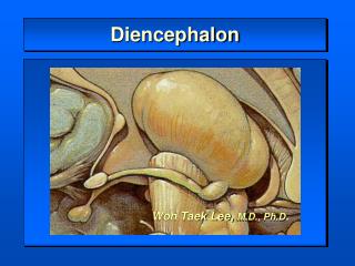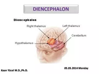
Diencephalon- Position
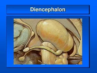
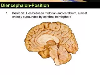
Diencephalon- Position. Position : Lies between midbrian and cerebrum, almost entirely surrounded by cerebral hemisphere. Diencephalon- Position.

Diencephalon- Position
E N D
Presentation Transcript
Diencephalon-Position • Position: Lies between midbrian and cerebrum, almost entirely surrounded by cerebral hemisphere
Diencephalon-Position The diencephalon consists of the hypothalamus, subthalamus, dorsal thalamus and epithalmus. These structures basically surround the third ventricle, and comprise the lateral wall and floor of this ventricle.
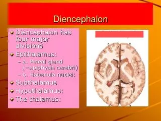
Subdivisions • Doral thalamus • Metathalamus • Epithalamus • Subthalamus • Hypothalamus
Thalamus The thalamus (from Greek "inner chamber") is a midline symmetrical structure within the brains of vertebrates including humans, situated between the cerebral cortex and midbrain. Its function includes relaying sensory and motor signals to the cerebral cortex, along with the regulation of consciousness, sleep, and alertness. The thalamus surrounds the third ventricle. It is the main product of the embryonic diencephalon.
Subdivisions DIENCEPHALON PARTS STRUCTURE FUNCTION
Dorsal thalamus External features • A large egg-shaped nucleus mass, • Anterior end called anterior thalamic tubercle, • Posterior end called pulvinar • Right and left portion of thalamus are joined by interthalamic adhesion • Floor-hypothalamic sulcus
Classification of nuclei of dorsal thalamus Three nuclear group-divided by internal medullary lamina • Anterior nuclear group • Medial nuclear group • Lateral nuclear group
Classification of Nuclei of DorsalThalamus internal medullary lamina Med. nuclear group Dorsal tier Ant. nuclear group Pulvinar Ventral anterior Medial geniculate body (MGN) Ventral intermediate Ventral posterior nucleus (VP) Lateral geniculate body (LGN) Ventral posterolateral (VPL) Ventral posteromedial (VPM )
Functional subdivision Nonspecific relay nuclei-receive afferents from rhinencephalon and reticular formation of brain stem, project mainly to hypothalamus and corpus striatum • Midline nucleus group • Intralaminar nuclear group • Thalamic reticular nucleus Association nuclei -receive input from many converging sours and in turn project widely to the association areas of cerebral cortex • Anterior nuclear group • Medial nuclear group • Dorsal tier of lateral nuclear group
Thalamic Nuclei Special relay nuclei • Vent. anterior nucleus (VA) • Vent. intermediate nucleus (VI) Receiving dentate nucleus, globus pallidus and substantia nigra to motor cortex • Vent. posteromedial nucleus (VPM)Receives trigeminal lemniscus and taste fibers • Vent. posterolateral nucleus (VPL)Receives medial lemniscus and spinal lemniscus Projects to first somatic sensory area via central thalamic radiation
Metathalamus Lateral geniculate body (LGN) Medial geniculate body (MGN) Metathalamus
Metathalamus • Medial geniculate body (MGN) • Relay station of audition • Receive fibers from inferior colliculus • Projects to auditory area via acoustic radiation • Lateralgeniculate body(LGN) • Relay station of vision • Receive fibers from optic tract • Projects to visual area via optic radiation
Epithalamus Includes • Thalamic medullary stria • Habenular trigone • Habenular commissure • Pineal body • Posterior commissure
Epithalamus • The epithalamus consists of the pineal gland and habenular nuclei. • The pineal is an interesting area containing modified photoreceptor cells. • These release melatonin in a circadian rhythm associated with the day/night cycle. • Despite the fact that pinealocytes are modified photoreceptors, they do not normally respond directly to light in humans. • They respond to circadian differences in sympathetic nervous system activity. • This may be important in sleep-wake cycles and, in many species it is the critical trigger for hibernation and sexual maturation.
Epithalamus • The habenular nuclei receive input from several limbic structures (including the ventral forebrain and septal nuclei) via the stria medullaris thalami. • This tract can be seen on the third ventricular surface of the thalamus. • The habenular nuclei project to the interpeduncular nucleus of the midbrain via the fasciculus retroflexus. • This is a pathway by which limbic structures may influence brain stem (reticular formation) function.
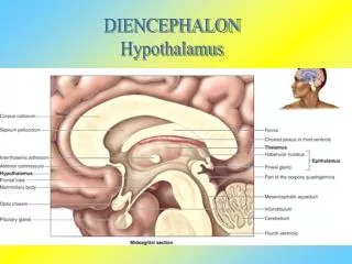
Position-lies ventral to thalamus Boundaries Superiorly: hypothalamic sulcus Inferiorly: Optic chiasma Tuber cinereum Infundibulum Mamillary body Anterior: lamina terminalis Posterior: continues with midbrain tegmentum Hypothalamus
Subthalamus • Transition zone between diencephalons and tegmentum of midbrain • Contain subthalamic nucleus, parts of red nucleus and substantia nigra
Subdivisions • Preoptic region • Supraoptic region • Tuberal region • Mamillary region
Important Nuclei • Supraoptic region • Supraoptic nucleus-produce antidiuretic hormone (ADH,vasopressin) • Paraventricular nucleus -produce oxytocin • Tuberal region • Infundibular nucleus • Ventromedial nucleus • Dorsomedial nucleus • Mamillary region • Mamillary nucleus • Posterior hypothalamic nucleus
Hypothalamic Nuclei Paraventricular nucleus Paraventriculohypophyseal tract Supraoptic nucleus Mamillary nucleus Supraopticohypophyseal tract Arcuate nucleus Tuberoinfundibular tract Infundibulum Anterior lobe of hypophsis Posterior lobe of hypophysis
Hypothalamus --connection • Connects with limbic system • Connects with brainstem and spinal cord • Connects with dorsal thalamus • Connects with hypophysis
Hypothalamic Connections • Supraoptic nucleus →supraoptic nucleus (ADH) →supraopticohypophyseal tract →posterior lobe of hypophysis • Paraventricular nucleus→ paraventicular nucleus (oxytocin) →paraventriculohypophyseal tract→posterior lobe of hypophysis
Hypothalamic Nuclei Paraventricular nucleus Paraventriculohypophyseal tract Supraoptic nucleus Supraopticohypophyseal trac Inferior hypophyseal a. posterior lobe of hypophysis Hypophyseal v.
Pituitary Gland Parvicellular neurons in the arcuate nucleus and nearby region of the walls of the third ventricle secrete releasing and inhibiting hormones → tuberoinfundibular tract →portal vein of hypophsis → anterior lobe of hypophsis Tuberoinfundibular tract Median eminence Portal v. Superior hypophyseal a. anterior lobe Hypophyseal v.
Hypothalamus Function • Regulates functions of neuroendocrine system • Autonomic nervous system
Third ventricle • Position: a narrow ventricle cleft lies within diencephalons • Boundaries • Roof: choroids plexus • Floor: optic chiasma, tuber cinereum, infundibulum and mamillary body • Anterior: lamina terminalis • Posterior: continuous with mesencephalic aqueduct • Lateral wall: dorsal thalamus and hypothalamus • Communication Third ventricle →mesencephalic aqueduct → fourth ventricle
Diencephalon • Relay between the brainstem & cerebral cortex • Dorsal-posterior structures • Epithalamus • Habenular nuclei – integrate smell & emotions • Pineal gland – monitors diurnal / nocturnal rhythm • Thalamus • Metathalamus • Medial geniculate body – auditory relay • Lateral geniculate body – visual relay • Ventral-anterior structure • Hypothalamus
Function of the Thalamus • Sensory relay • ALL sensory information (except smell) • Motor integration • Input from cortex, cerebellum and basal ganglia • Arousal • Part of reticular activating system • Pain modulation • All nociceptive information • Memory & behavior • Lesions are disruptive
Input to the Thalamus Metathalamus Vision and Hearing
Input to the Thalamus Sensory relay - Ventral posterior group all sensation frombody and head, including pain
Input to the Thalamus Motor control and integration
Input to the Thalamus Behavior and emotionconnection with hypothalamus
Projections from the Thalamus Metathalamus Vision and Hearing
Projections from the Thalamus Sensory relay Ventral posterior group all sensation frombody and head, including pain
Projections from the Thalamus Motor control and integration