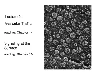
Vesicular Traffic II
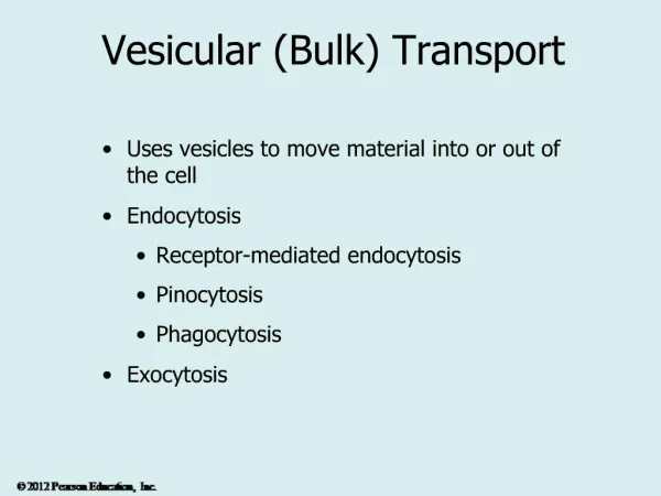
Vesicular Traffic II. Endocytic and secretory pathways. red = secretory green = endocytic blue = recycling. Different coats are used for different transport steps in the cell. Assembly and disassembly of clathrin coat. Dynamin pinches clathrin coated vesicles from the membrane.

Vesicular Traffic II
E N D
Presentation Transcript
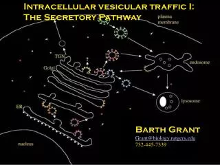
Endocytic and secretory pathways red = secretory green = endocytic blue = recycling
Different coats are used for different transport steps in the cell
Interaction of SNARE proteins while docking synaptic vesicle at nerve terminal
Model for membrane fusion. Tight SNARE pairing forces water molecules from area between lipid bilayers
Vesicular tubular clusters move along microtubules to carry proteins from ER to Golgi apparatus
Some enzymes are enriched in cis or trans compartments of the Golgi substrates specific for certain enzymes such as acid phosphatase
Golgi apparatus can be polarly distributedIn this fibroblast Golgi is facing the direction in which the cell is crawling
Drawing of goblet cell in the intestinal epithelium Secretes polysaccharide rich mucus into the small intestine Golgi is highly polarized to facilitate release of mucus through exocytosis at the apical domain
N-linked glycosylation because sugar is added to N of asparagine. original precursor oligosaccharide added to most proteins in the ER
Oligosaccharide chains are processed in the Golgi complex common core high-mannose
Detection of acid phosphatase in lysosomesSmall spheres may be vesicles delivering the enzyme from the Golgi Apparatus