Download
1 / 75
830 likes | 1.13k Vues
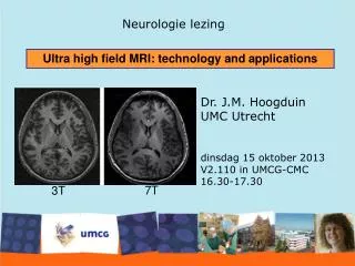
Explore the evolution of MRI technology with the rise of ultra-high-field strengths, such as 7T, for improved diagnostic accuracy, shorter acquisition times, and enhanced clinical throughput. Discover how these advancements have revolutionized imaging techniques and spectroscopy in both human and animal models.

E N D
Ultra High Field MRI Charles Dietz MD 2017
4 TESLA (1991) MDEFT Barfuss et.al. NMR Biomed:3(1)1990 (DATA from SIEMENS) SIEMENS, GE, and PHILIPS
1991 4 Tesla CMRR BOLD fMRI a R Full Field Visual Stimulation b HEMI Field Visual Stimulation c Ogawa et al. Proc Natl Acad Sci USA (1992) 89, 5951-5955
1 2 3 Functional Imaging with Magnetic Resonance Visual Stimulus 4 Tesla Title Ogawa et al. Proc Natl Acad Sci USA (1992) 89, 5951-5955
0.2% 4% R L R L Wei Chen et al., U of Minnesota, CMRR, 4T
Opaque anatomical Image In Grey scale SILENT WORD GENERATION
DEVELOPMENT OF CMRR INSTRUMENTATION for HIGH MAGNETIC FIELD IMAGING and SPECTROSCOPY 4.7 Tesla/40 cm (~1985) • 4 Tesla Human (1991) 9.4 Tesla/31 cm (~ 1995) • 7 Tesla Human (~1999) 3 Tesla ~2002 • 9.4 Tesla Human (~2005) 2nd 7T (2010) and 10.5T (2015) 16.4 Tesla /26 cm bore (animal) (2010)
7T MRI Department of Radiology Charles Dietz
7 Tesla/90 cm bore ~1999
7 Tesla UHF Clinical MRI • Ultra Highfield (UHF) MRI at 7T with increased S/N ratio • Spectroscopy • Higher Resolution – Improved Diagnostic Accuracy • Shorter Acquisition Time – Higher Clinical Throughput
Patellar Cartilage 7T 3T
Meniscal Tears 7T 3T
Knee imaging at 7T a) 16-element transmit/receive for all knee images. b) Sagittal turbo spin echo (TSE) image (TR/TE = 4210/31 ms, resolution 0.2x0.2x3, 39 slices, 1 average, echotrain length 7, 10 min. c) Axial turbo spin echo (TSE) image (TR/TE = 4680/27 ms, resolution 0.3x0.3x2, 29 slices, 2 averages. Ellermann et al. DETAILED ANATOMY OF TIBIAL NERVE AT 7T (CMRR)
Quantitative cartilage assessment: T1ρ-maps of articular cartilage Dog model of early osteoathritis at 7 Tesla (CMRR) T1ρ Surgery Control preparationmodule nx4 hyperbolic secant (HS1)-pulses pulse duration: 5.12 ms imaging module in-plane resolution: 620 μm slice thickness: 1.5 mm matrix: 128×128 # segments: 4 TE/TR = 5.1 ms/4 s T1ρ of the cartilage in the severed joint is longer than in the control Pepin SR et al: A comparative analysis of 7.0-Tesla magnetic resonance imaging and histology measurements of knee articular cartilage in a canine posterolateral knee injury model: a preliminary analysis. Am J Sports Med. 2009;37 Suppl 1:119S-24S.
IMAGING AT HIGH AND ULTRAHIGH FIELDS IN THE HUMAN TORSO; THEN (1990; 4 TESLA) 4 Tesla (Data from Siemens) Barfuss et al. NMR Biomed:3(1)1990 NOW (7 TESLA) 7 Tesla Henry, TR et al., Radiology 2011;261(1):199-209.
7 Tesla (CMRR) Snyder, C. et al MRM 61(3):517-524 (2009) IMAGING AT HIGH AND ULTRAHIGH FIELDS IN THE HUMAN TORSO; THEN (1990; 4 TESLA) 4 Tesla (Data from Siemens) Barfuss et al. NMR Biomed:3(1)1990 NOW (2009; 7 TESLA)
7 TESLA: A 16-channel combined loop-dipole transceiver array for 7 Tesla body MRI Ertürk et al. MRM (2016). 26
STN SN RN Anatomical contrast @ 7T SWI T1W T2W SWI: Susceptibility-weighted imaging STN: subthalamic nucleus SN: substantia nigra RN: red nucleus Abosch, Yacoub, Ugurbil, Harel. 2010
Susceptibility-Weighted Imaging @ 7T STN SN STN = Subthalamic Nucleus SN = Substantia Nigra Magnet: 7T Resolution: 0.4 x 0.4 x 0.8 mm Dr. Noam Harel, University of Minnesota / CMRR
Susceptibility-Weighted Imaging @ 7T Schaltenbrand and Wahren Atlas (Ex-vivo) SWI @ 7T (In-vivo) Lamina pallidi medialis GPe GPe GPi GPi GP = Globus pallidus Abosch, Yacoub, Ugurbil, Harel. 2010
DBS STN SN RedN Dr. Noam Harel, University of Minnesota / CMRR
Left Orientation Right Phase 1 mm Yacoub, Harel, Uğurbil PNAS (2008) 105(30): 10607-12 ODC 1 mm Yacoub, Shmuel, et al. Neuroimage (2007) 37(4): 1161-77
Tonotopic Mapping in Human Primary Auditory Cortex 7 T GE fMRI 1.2x1.5x2.4 mm3 CMRR/U Maastricht
Laminar Resolution De Martino et al (PNAS 2015) Medial Geniculate Body Inferior Colliculus Moerel et al (Scien.Rep.,5; 17048 (2015)) De Martino et al. (Nature Communications, 2013) High Resolution Frequency maps (tonotopy) in the Human Brain at 7 Tesla Primary Auditory Cortex Formisano et al. (Neuron, 2003)
Zimmermann et al. PLoS ONE 6(12): e28716. (2011) MT Flattened maps of 3 layers surface Zimmermann et al. PLoS ONE 6(12): e28716. (2011) CMRR & Univ. Maastricht Collaboration deep 35 0.8 mm isotropic
NAA Cr PCr Cr PCr scyllo-Ins Glu Gln Glu Cho GSH Tau Ins Gln NAA GABA Ins residual water Asp Glc 7 TESLA HUMAN BRAIN 1H NMR spectrum at STEAM TE = 6 ms TR = 5 s VOI = 8 ml NT = 160 36
lactate: resonates at 1.3 ppm lipids: resonates at 1.3 ppm alanine: resonates at 1.48 ppm N-acetylaspartate (NAA): resonates at 2.0 glutamine/glutamate: resonates at 2.2-2.4 ppm GABA: resonates at 2.2-2.4 ppm 2-hydroxyglutarate: resonates at 2.25 ppm 6 citrate: resonates 2.6 ppm creatine: resonates at 3.0 ppm choline: resonates at 3.2 ppm myo-inositol: resonates at 3.5 ppm water resonates at 4.4 ppm 37
7T, STEAM TR/TE=5000/6 ms VOI=20 x 22 x 20 mm3 32 scans During visual stimuli, changes of metabolite concentrations were within ± 0.2 mmol/g 38
Results - 5.3 min stimulation Group Analysis Error bars = CRLB 39
SPINOCEREBELLAR ATAXIAS N-acetylaspartate (NAA): resonates at 2.0 40
GOAL: Develop the imaging hardware and methods to exploit the advantages of UHF imaging in the prostate. UHF Prostate • Development of novel RF coils • Surface arrays • Multi-channel endorectal Coils • Improved Imaging and Spectroscopy • Increased resolution anatomic imaging • Increased resolution functional imaging • Improved spectral quantification on spectroscopy • Future Work: evaluate the potential advantages in cancer detection and staging to improve the identification of clinically significant disease.
Sagittal T2 Coronal T2 Axial ADC Axial T2 3T 7T
7T Intro Our work on 7T (UHF) breast MR has focused on RF coil development. We’ve made 4 iterations of RF coils, and while we’ve used them to get some prelim data they are all unsatisfying. The problem is that for contrast-enhanced breast imaging you need relatively uniform flip angles over both breasts and axillae. With the reduced RF wavelength at 7T (~12cm) it is very difficult to achive this. This is why we’ve had more success with smaller regions (prostate, cardiac, kidneys) that are more tolerant to flip angle variation.
7T Breast MR Imaging High SNR allows high-resolution anatomic imaging Axial fat-sat 3D FLASH 0.5 x 0.5 x 2 mm Sagittal fat-sat 3D FLASH 0.3 x 0.3 x 2.2 mm
6 6 Normal Volunteer 4 Patient with IDC 4 2 2 0 0 ppm ppm Haddadin I. et al., NMR Biomed 2009 Bolan P. J. et al., ISMRM 2006 7T Breast MR Spectroscopy Increased spectral resolution and SNR gives excellent spectra in normals and patients choline: resonates at 3.2 ppm Bolan et al., ISMRM 2006
3T Advancements • The following slides are 3T only: they describe the development of high-resolution diffusion imaging
High-resolution Breast DWI: RS vs SMS-IPSE T2-weighted anatomical Standard b = 0 • Comparing approaches with fixed 5 min acquisition time • Std, single-shot EPI • RESOLVE (RS-EPI), following Wisner JMRI 2014 • SMS/MB with in-plane slice encoding 0.8 x 0.8 x 3 mm 1.7 x 1.7 x 4 mm RS-EPI b = 0 SMS/MB-IPSE b = 0 1.8 x 1.8 x 2.4 mm 1.25 x 1.25 x 2.5 mm SMS/MB-IPSE approaches T2 resolution & quality
400 (mL/100 g/min) Renal Blood Flow 0 Li, NMR in Biomed:63-69; 2015 Li (2015) ISMRM #3155.
NIH NIH invests substantially (~7-8 billions/year) in Neurosciences and Brain disorders Research Brain Research through Advancing Innovative Neurotechnologies (BRAIN) Initiative 49
Description of the functional and structural connections among gray matter locations in the human brain • spontaneous fluctuations in an fMRI time series (i.e. “Resting State” fMRI ) to deduce ‘functional connectivity’ and/or • Diffusion weighted MRI to infer ‘structural connectivity’ 50