Download
1 / 28
280 likes | 522 Vues
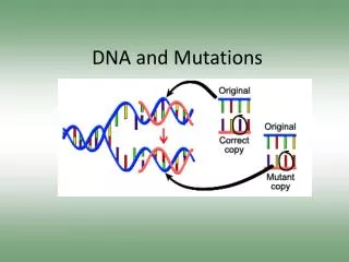
Mutations and Karyotyping. Mutations Ch. 10. pg. 219-220. Changes in nucleotide sequence of DNA May occur in somatic cells (body cells) not passed to offspring May occur in gametes (eggs & sperm) passed to offspring. Causes. Mutations happen regularly

E N D
Mutations Ch. 10. pg. 219-220 • Changes in nucleotide sequence of DNA • May occur in somatic cells (body cells) • not passed to offspring • May occur in gametes (eggs & sperm) • passed to offspring
Causes • Mutations happen regularly • Any agent that causes a change in DNA is called a mutagen. • Mutagens include radiation, chemicals, and even high temperatures. • Ex. of radiation: X rays, cosmic rays, ultraviolet light, and nuclear radiation. • Chemicals: Benzene • Many mutationsare repaired by enzymes.
Are Mutations Helpful or Harmful? • Some harmful - Skin cancers and some leukemias result from somaticmutations • Most mutations have no affect, some have detrimental affects and a few mutations may improve an organism’s survival (beneficial)
Types of Mutations • 2-Types • 1. Gene mutations - change in one DNA sequence of a gene. • 2.Chromosomal mutations – change in structure or loss or gain of part of a chromosome.
Gene mutation- 2 types1. Point Mutation • A change in a single base pair in DNA. • Changes the amino acidin the protein • Does not always cause a problem. • THE DOG BIT THE CAT • THE DOG BIT THE CAR
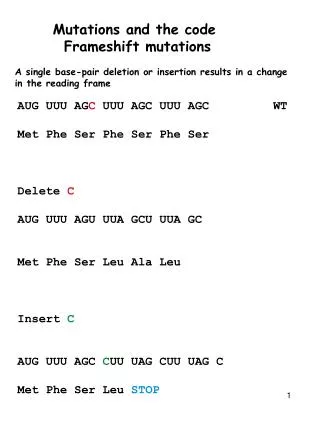
2. Frameshift Mutation • A single base is added or deleted from the DNA causing all the other bases to be out of position. • More harmful than a regular point mutation. • THE DOG BIT THE CAT • THE DOB ITT HEC AT
Chromosome Mutations • Five types exist: • Deletion • Inversion • Translocation • Nondisjunction • Duplication
DeletionPartof a chromosome is lost/deleted. ABCDEFGH A B C E F G H
Duplication/InsertionA part of the chromosome repeats A B C D E F G H A B C B C D E F G H
InversionPart of a chromosome breaks off;reattaches backward A B C D E F G H H A D C B E F G
TranslocationPart of a chromosome breaks off; attaches to a different chromosome that is not homologous A F G H E B C D F G W X B C D E H A X Y Z Y Z W X Y Z Y Z W Translocation Translocation
Nondisjunction • Failureof pair of chromosomes to separate during meiosis • Example Down Syndrome
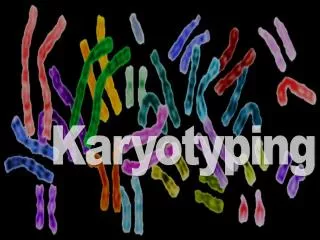
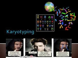
Karyotyping Ch. 6 pg. 122-123
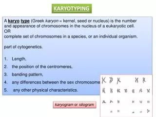
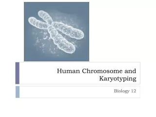
Some mutations that cause chromosomal abnormalities can be detected by analyzing a karyotype. • Karyotype – photo of the chromosomes in a dividing cell that shows the chromosomes arranged by size, number, and shape. • Identifies gender and genetic disorders
Amniocentesis – medical procedure used in prenatal diagnosis of chromosomal abnormalities and fetal infections • small amount of amniotic fluid, which has fetal tissues, is extracted from the amniotic sac surrounding a developing fetus • 2 types of abnormalities in chromosomes • Autosomal abnormalities– abnormalities of chromosomes not directly involved in determining gender • Sex Chromosomal abnormalities– abnormalities that affect the gender of an individual • XX – female • XY - male
How Scientists Read Chromosomes? • Size. This is the easiest way to tell two different chromosomes apart. • Banding pattern. The size and location of Giemsa bands on chromosomes make each chromosome pair unique. • Centromere position. Centromeres are regions in chromosomes that appear as a constriction. They have a special role in the separation of chromosomes into daughter cells during mitosis cell division (mitosis and meiosis). To "read" a set of human chromosomes, scientists first use three key features to identify their similarities and differences: Image taken from: http://learn.genetics.utah.edu/content/begin/traits/scientists/ Using these key features, scientists match up the 23 pairs – one set from the mother and one set from the father.
Trisomy 21- Down’s Syndrome - Karyotype 47 Total Chromosomes Three Chromosomes at the 21st Pair Image taken from: http://worms.zoology.wisc.edu/zooweb/Phelps/karyotype.html
Monosomy X - Turner Syndrome - Karyotype 45 Total Chromosomes One “X” Chromosome Image taken from: http://worms.zoology.wisc.edu/zooweb/Phelps/karyotype.html
XYY Karyotype 47 Total Chromosomes One “X” and Two “Y” Chromosomes Image taken from: http://worms.zoology.wisc.edu/zooweb/Phelps/karyotype.html
Examples Cri du chat – deletion of #5 Wilson’s Disease – can’t get rid of excess copper Down's syndrome – extra copy of #21 Patau Syndrome – extra copy of #13 Klinefelter's syndrome (XXY) Turner syndrome (X instead of XX or XY) XYY syndrome. XYY Triple-X syndrome (XXX) XXXX Syndrome
Colored Blindness Hemophilia – bleeding disorder Huntington’s Disease – faulty protein (mental/nervous disorder) Jackson-Weiss Syndrome – fusion of face/foot bones Sickle Cell Disease – red blood cells sickle shaped Tay-Sachs Disease – brain/nerve disorder Polydactyly – extra digit