
Chromosomes and Karyotyping
E N D
Presentation Transcript
1. Chromosomes and Karyotyping
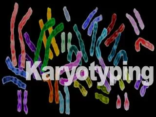
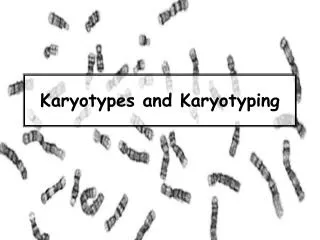
2. Chromosome Structure Chromosomes under a microscope stained with dye
Striped rod-shaped structure
3. Chromosomes are found in the nucleus
Chromosomes are made of a DNA strand that is tightly wound around itself and packed with protein called histones
The �stripes� observed are a result of the dyes binding to the specific proteins
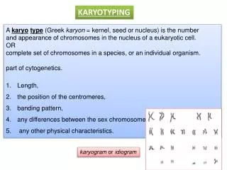
Each chromosome pair has a unique banding pattern. Chromosome Structure
4. Chromosome Structure During Mitosis, Chromosomes Replicate (full copy)
Replicated chromosomes attach at the centromere
Called sister chromotids
5. Chromosome Structure Humans have 23 different chromosomes
Humans have 2 copies of each chromosome
Diploid = 2 copies
Total of 46 chromosomes in EVERY Cell.
Chromosome partners are called homologous chromosomes
6. Seeing Chromosomes Joe Hin Tijo and Albert Levan�1956
Developed approach to visualize chromosomes: �Catching Chromosomes�
Chromosomes condense during mitosis
Treat cell with a drug to stop cell division
Squash the cells and view under microscope
Photograph the microscope picture
Clip the picture and organize the chromosomes into a karyotype