Download
1 / 68
680 likes | 821 Vues
Imaging the normal and abnormal lung There is minimal descriptive text in the presenter’s notes field Please email me if you have questions Dr I Runcie Consultant Radiologist PRH Ian.runcie@bsuh.nhs.uk. http://alberich4.tripod.com/history/100years.html. 2. Chest X-ray CT scan

E N D
Imaging the normal and abnormal lung There is minimal descriptive text in the presenter’s notes field Please email me if you have questions Dr I Runcie Consultant Radiologist PRH Ian.runcie@bsuh.nhs.uk
Chest X-ray • CT scan • Ventilation and perfusion imaging
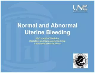
This is a radiograph of what appears to be a bunch of grapes. Would you / could you eat these grapes?
The radiologist’s radiographic world view Water - all body fluids and tissues except fat and bone Fat Gas High atomic number calcium iodine barium metals
Pulmonary arteries ✓ Pulmonary veins ✓ Bronchi ?
CT scan of the lungs • Shows amazing detail • Resolve small structures • Resolve small differences in attenuation
CT thorax and HRCT lung • Slice thickness • Resolution • Thin section (1mm) is the optimal technique for demonstrating lung • CT thorax 5-8mm slice thickness
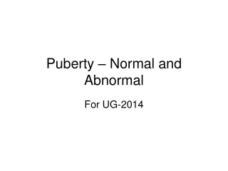
2 4 1 4 5 3
Structure and function The pulmonary lobule and acinus are functional units of the lung. Secondary pulmonary lobule 5-20mm dia Acinus 6-10mm - 300M alveoli per adult – total area 143m2
2 1
What are lungs for? How could we image function?
Lung function - Gas exchange Ventilation - move air in and out Perfusion - move blood in and out Diffusion - moves gases between alveoli and blood
How could we obtain a picture of the distribution of ventilation in the lungs?
Ventilation Radioactive gas - Xenon-133, Krypton-81m Radioactive aerosol - Tc99m-DTPA, Radioactive dry carbon particles 20nm (0.02 microns) 50micrograms Tc99m (2 0rders of magnitude below 24 hour permitted atmospheric pollution)
How could we produce a steady state image of lung blood flow?
If you inject particles into a peripheral vein where will they stop? Does this carry any risk?
Lung perfusion 2-500,000 intravenous particles 16-90 micron diameter Macroaggregated human serum albumen Occlude <1% of pulmonary circulation
What patient posture would achieve the most uniform distribution of particles throughout the lungs? Upright? Lying on the right side? Lying flat on back?
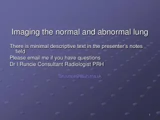
R What has happened to the right lung
What are the round white objects in the lower part of each lung field?
Lung imaging A danger of DIY How is the patient breathing? What might her blood gases be like?
Air space shadowing = consolidation • Fluffy blobs • About 1cm dia • Tending to confluence • Could be oedema /transudate / exudate / pus / blood • +/- air bronchogram