Lung Examination: Abnormal
LOYOLA UNIVERSITY MEDICAL CENTER Loyola University Chicago. Lung Examination: Abnormal. Arcot J. Chandrasekhar, M.D. December 1, 2009. Respiratory System. Lungs Airways Pleura Mediastinum Chest Wall Respiratory Centers. Pathological Correlation. Localized Disease Consolidation

Lung Examination: Abnormal
E N D
Presentation Transcript
LOYOLA UNIVERSITY MEDICAL CENTER Loyola University Chicago Lung Examination: Abnormal Arcot J. Chandrasekhar, M.D. December 1, 2009
Respiratory System • Lungs • Airways • Pleura • Mediastinum • Chest Wall • Respiratory Centers
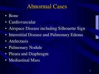
Pathological Correlation • Localized Disease • Consolidation • Cavitation • Mass • Atelectasis • Pleural Disease • Pleural effusion • Pneumothorax • Diffuse Lung Disease • Emphysema • Diffuse airway disease • Diffuse alveolar disease • Diffuse interstitial disease • Mediastinal Disease • Respiratory Centers
Physical Exam Steps • General examination • Mediastinal position • Chest expansion • Lung resonance • Breath sounds • Adventitious sounds • Voice transmission
General Examination • Respiratory rate • Pattern of breathing • Cyanosis • Clubbing • Weight • Cough • Hospital setting • Effort of ventilation • Shape of thorax
Respiratory Rate • Bradypnea: rate less than 8 per minute • Tachypnea: rate greater than 25 per minute
Pattern of Breathing • Kussmals • Sleep apnea • Cheyne strokes • Pursed lip breathing • Orthopnoea: Short of breath in supine position, gets some relief by sitting or standing up
Central Cyanosis • Results from pulmonary dysfunction, the mucous membrane of conjunctiva and tongue are bluish. • If there was chronic hypoxemia and secondary erythrocytosis, you can detect the conjunctival and scleral vessels to be full, tortuous and bluish.
Clubbing • In clubbing, there is widening of the AP and lateral diameter of terminal portion of fingers and toes giving the appearance of clubbing. • The angle between the nail and skin is greater than 180. • The periungual skin is stretched and shiny. • There is fluctuation of the nail bed. • One can feel the posterior edge of the nail.
Significance: Clubbing Observed In: • Intrathoracic malignancy: Primary or secondary (lung, pleural, mediastinal) • Suppurative lung disease: (lung abscess, bronchiectasis, empyema) • Diffuse interstitial fibrosis: Alveolar capillary block syndrome • In association with other systemic disorders
Weight • Emaciation cachectic • Malignancy • Tuberculosis
Weight • Obese: Sleep apnea syndrome
Cough • Productive • Dry • Whooping • Bovine
Hospital Setting • Isolation room • Oxygen set up
Effort of Ventilation • Patient appears uncomfortable. Breathing seems voluntary. • Accessory muscles are in use, expiratory muscles are active and expiration is not passive any more. • The degree of negative pleural pressure is high. • The respiratory rate is increased.
Resting Size and Shape of Thorax • Barrel chest • Kyphosis • Scoliosis • Pectus excavatum • Gibbus
Barrel Chest AP Diameter = Transverse Diameter
Tracheal Position: Mediastinum • Any deviation of the mediastinum is abnormal • Lateral shift: The mediastinum can be either pulled or pushed away from the lesion • Pull: Loss of lung volume (Atelectasis, fibrosis, agenesis, surgical resection, pleural fibrosis) • Push: Space occupying lesions (pleural effusion, pneumothorax, large mass lesions) • Mediastinal masses and thyroid tumors
Chest Expansion • Asymmetrical chest expansion is abnormal • The abnormal side expands less and lags behind the normal side • Any form of unilateral lung or pleural disease can cause asymmetry of chest expansion • Global expansion decrease
Percussion: Decreased or Increased Resonance is Abnormal • Dullness • Decreased resonance is noted with pleural effusion and all other lung diseases • The dullness is flat and the finger is painful to percussion with pleural effusion • Hyper resonance: Increased resonance can be noted either due to lung distention as seen in asthma, emphysema, bullous disease or due to Pneumothorax • Traube's space
Breath Sounds: Diminished or Absent • Intensity of breath sounds, in general, is a good index of ventilation of the underlying lung. • Breath sounds are markedly decreased in emphysema. • Symmetry: If there is asymmetry in intensity, the side where there is decreased intensity is abnormal. • Any form of pleural or pulmonary disease can give rise to decreased intensity. • Harsh or increased: If the intensity increases there is more ventilation and vice versa.
Bronchial • Bronchial breathing anywhere other than over the trachea, right clavicle or right inter-scapular space is abnormal. • In consolidation, the bronchial breathing is low pitched and sticky and is termed tubular type of bronchial breathing. • In cavitary disease, it is high pitched and hollow and is called cavernous breathing. You can simulate this sound by blowing over an empty coke bottle.
Rhonchi • Rhonchi are long continuous adventitious sounds, generated by obstruction to airways. • When detected, note whether it is generalized or localized, during inspiration or expiration, and the pitch. • Diffused rhonchi would suggest a disease with generalized airway obstruction like asthma or COPD.
Rhonchi Asthmatic Continuous
Rhonchi • Localized rhonchi suggests obstruction of any etiology e.g., tumor, foreign body or mucous. • Mucous secretions will disappear with coughing, so would the rhonchus. • Expiratory rhonchi implies obstruction to intrathoracic airways. • Asthmatics can also have inspiratory rhonchi while it is uncommon in COPD.
Pleural Rub • Normal parietal and visceral pleura glide smoothly during respiration. • If the pleura is roughened due to any reason, a scratching, grating sound, related to respiration is heard. • You can hear the sound by compressing harder with the stethoscope and making the patient take deep breaths. • It is localized and can be palpable.
Pleural rub Scratching, Grating Related to respiration
Stridor • Loud audible inspiratory rhonchi is called a stridor. • Inspiratory rhonchi in general, implies large airway obstruction.