F. Sensory Receptors
670 likes | 2.67k Vues
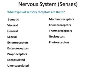
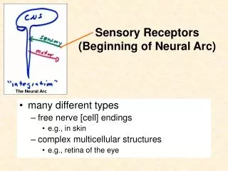
F. Sensory Receptors. What are sensory receptors? http://www.youtube.com/watch?v=1vLsZ_dXFAg&feature=related a life without pain. Sensory Receptors are: highly modified ends of sensory neurons that are activated by a specific stimulus

F. Sensory Receptors
E N D
Presentation Transcript
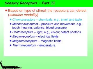
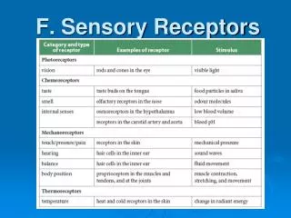
What are sensory receptors?http://www.youtube.com/watch?v=1vLsZ_dXFAg&feature=relateda life without pain Sensory Receptors are: • highly modified ends of sensory neurons that are activated by a specific stimulus • specialized cells that receive stimuli and translate them to a nerve message in a sensory neuron • Sensory adaptation occurs once the receptor has become accustomed to the stimulus. The neuron ceases to fire even though the stimulus is still present (often occurs with the sense of smell).
Taste One cannibal to another while eating a clown: "Does this taste funny to you?" • there are 300 taste buds on the tongue • the taste is perceived in the parietal lobe of the brain • the tongue has four different taste receptors (salty, sweet, sour and bitter) See fig. 12.25
http://www.vivo.colostate.edu/hbooks/pathphys/digestion/pregastric/taste.htmlhttp://www.vivo.colostate.edu/hbooks/pathphys/digestion/pregastric/taste.html http://www.agen.ufl.edu/~chyn/age2062/lect/lect_24/lecture_24.htm
Smell • olfactory (smell) cells are located in the nasal cavity • airborne chemicals combine with the receptor ends on olfactory cells to create an action potential • the smell is perceived by the temporal lobe of the brain • the senses of taste and smell work together
http://instruct1.cit.cornell.edu/courses/biog105/pages/demos/105/unit10/tastesmell.htmlhttp://instruct1.cit.cornell.edu/courses/biog105/pages/demos/105/unit10/tastesmell.html
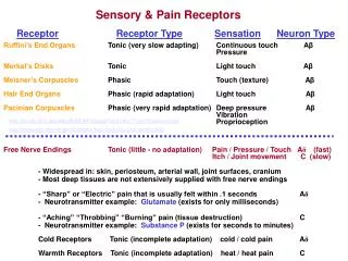
2. Pressure • a mechanical stimulus involving the movement of the skin or changes in the body surface • Includes touch and pain
3. Proprioreceptors • a mechanical stimulus involving movement of the muscles, tendons and joints in the arms and/or legs • Responsible for maintaining body position
4. Heat and cold • thermoreceptors detect a change in temperature on the surface of the skin
5. Balance • body movement equilibrium is maintained by specialized structures in the inner ear • Fluid movement causes the movement of special hair cells in the inner ear, which results in a stimulus and eventual response
a. Structureshttp://www.youtube.com/watch?v=Sqr6LKIR2b8 • sclera (outer eye) – the white portion of the eye, supports and protects photocells • cornea (outer eye) – transparent tissue that bends light toward the pupil. Requires oxygen and nutrients but has no blood vessels so most of the oxygen absorbed from the atmosphere are found in tears • lens (accessory) – focuses the image on the retina, flatten/narrows as you focus near or far • iris (middle eye) – regulates the amount of light entering the eye
a. Structures • pupil (middle eye) – hole in the iris, allows light to penetrate the eye • aqueous humor (accessory) – supplies the cornea with nutrients and bends light entering the eye • vitreous humor (accessory) – holds retina and lens in place • retina (inner eye) – contains photoreceptors that respond to light • fovea centralis (inner eye) –sensitive area of the retina, contains only cones
a. Structures • blind spot (inner eye) – area where the optic nerve attaches to the retina • choroid layer (middle eye) – contains pigments that prevent the scattering of light in the eye by absorbing stray light – contains blood vessels • optic nerve (accessory) – carries impulses from the rods and cones to CNS • rods – used for seeing in dim light • cones – identify color, used for sharp vision
b. Focusing image • light passes through the cornea, lens and fluid humours where it is bent • the image is sent to the fovea centralis of the retina where it if fixed smaller, upside down and reversed from left to right • when nearby objects are viewed, the ciliary muscles contract causing the lens to become rounder • when far-away objects are viewed, the ciliary muscles relax causing the lens to become flattened • the ability of the lens to change shape in order to focus images clearly on the retina is a reflex called accommodation (6 m away requires no accommodation)
c. Chemistry of vision • vision begins as light is focused on the light-receiving cells called photoreceptors (rods and cones)
Rods • rods are responsible for detecting motion and peripheral vision • rods are spread throughout the retina but are more concentrated on the outside edges • rods contain a pigment called rhodopsin, made of Vitamin A (retinal) and a protein called opsin http://health.howstuffworks.com/eye2.htm
when a small unit of light strikes the rhodopsin molecule it splits into retinal (pigment) and opsin (protein). • A cascade of reactions result in the closure of ion channels in the rod cell’s plasma membrane • Inhibition of an inhibitory transmitter causes an action potential to travel to the visual areas of the cerebral cortex • A signal is transmitted to the optic nerve
in bright light, rhodopsin is broken down faster than it can be restored • rods incapacitated until they can be restored • in the absence of light, retinal is changed to Vitamin A, and with the addition of ATP, the Vitamin A recombines with opsin to restore rhodopsin levels • nocturnal animals have a higher ratio of rods to cones • Rods allow for vision at night
Cones • cones allow for colour vision • vitamin A combines with three different protein opsins in the cones, each sensitive to the three primary colors of light (red, blue, green) • each pigment is located in a different cone http://health.howstuffworks.com/eye2.htm
d. Visual Interpretation • image is sent to the brain via stimulation of photoreceptor cells (rods and cones) which generate an action potential via the optic nerve to the brain • Blind spot – located on the retina where the optic nerve exists, there are no photoreceptors present • the signal proceeds to the optic chiasma • Nerves from both eyes come together and some cross to opposite sides of the brain • allow for binocular vision (3-D) • left and right visual fields cross to opposite hemispheres (occipital lobe) • Signal enters the primary visual cortex (information is not interpreted) • visual association area interprets image information and flips/rotates image
http://www.youtube.com/watch?v=QALdBU670Ro http://www.benbest.com/science/anatmind/anatmd5.html http://cti.itc.virginia.edu/~psyc220/
d. Vision defects • glaucoma – increase in vitreous humor causes the pressure to collapse in the blood vessel in the retina, no nutrients and oxygen are available and blindness results • http://www.youtube.com/watch?v=cF0rj4fM1l0&feature=related http://www.springereye.com/glaucoma.html
cataract – lens/cornea becomes clouded due to the degeneration of protein structure http://www.prof-vision.com/cataracts.html
astigmatism – lens/cornea is irregular, focus is not sharp (light rays do not meet at the correct focal point) http://www.lasersurgeryforeyes.com/astigmatism.html
near sightedness (myopia) – the lens is elongated, the image focuses in front of retina, so it is difficult to see objects far away • - the lenses worn to correct myopia are concave http://hcd2.bupa.co.uk/fact_sheets/html/myopia.html
far sightedness (hyperopia) –the image focuses behind the retina, so it is difficult to see objects close up • - convex lenses correct hyperopia • Lasik surgery http://www.youtube.com/watch?v=a7VRghAtwXU • http://www.youtube.com/watch?v=GaoA4PLb7hc&feature=related • http://www.youtube.com/watch?v=7PJ391MDtpo&feature=related http://www.doctorergo.com/home.html?main=consumer/refractive.html
color blindness – occurs when one or more type of cone is defective (red-green), defect is genetic and occurs more often in males than in females http://www.eyecaresource.com/conditions/color-blindness/
a. Outer Ear • pinna (auricle)– the external ear flap collects sound • auditory canal – carries sound to the eardrum, is lined with specialized sweat glands (ceruminous glands) that produce earwax to trap foreign invaders • Tympanic membrane (eardrum) – membrane that converts sound waves into mechanical motion (amplifies the sound) and transmits the sound to the middle ear • Separates outer and middle ear http://www.entnet.org/healthinfo/ears/swimmers.cfm
b. Middle Ear • ossicles – tiny bones which amplify and carry sound (malleus/hammer, incus/anvil, stapes/stirrup) • oval window – receives sound waves from the ossicles. The stirrup touching on the membrane passes on to cochlea • Eustachian tube – air filled tube connected to the throat which equalizes pressure between inner and outer ear http://www.oticon.com/eprOtiScripts/Files/encyclopedia/dir.asp?selectedID=198
c. Inner Ear • vestibule – connected by oval window, concerned with static equilibrium (position of head, movement along one plane) • utricle and saccule establish head position • Contain otoliths which move due to force of gravity • Causes fluid in the vestibule to move and the stereocillia to bend • Action potential is sent via the auditory nerve • Brain adjusts body to maintain balance http://paperairplane.mit.edu/16.423J/Space/SBE/neurovestibular/NeuroVestibular/2_Physiology/PhysSub2.html
c. Inner Ear • semicircular canals – movement of fluids here help identify body movement (dynamic equilibrium) • The base of each canal has an ampulla • Inside the ampulla are stereocillia embedded in gelatinous material (endolymph) • Change in position causes the fluid to flow within the ampulla, resuting in the stereocillia bending • Nerve impulses relay information to the brain about body position and motion • http://www.youtube.com/watch?v=u84_BfK3hcw&feature=related • http://www.youtube.com/watch?v=mmBB2bu1gEQ&feature=related
c. Inner Ear • cochlea – used for hearing, this is where mechanical energy of sound is converted to electrochemical impulses that are transmitted to the brain • The inner ear is filled with fluid and vibrations in the oval window are converted to pressure waves in the fluid • The cochlea can be divided into the organ of Corti, the basillarmembreane and the tectorial membrane. • organ of Corti - the organ of hearing • basilar membrane – on the base of the organ of Corti, sensory mechanoreceptors called hair cells are found here • the hair cells have projections called stereocillia • the stereocillia are embedded in the tectorial membrane • round window – lets vibrations out and controls pressure in the cochlea
http://www.oticon.com/eprOtiScripts/Files/encyclopedia/dir.asp?selectedID=198http://www.oticon.com/eprOtiScripts/Files/encyclopedia/dir.asp?selectedID=198
d. Hearing • sound waves push against eardrum • vibrations are passed on to three bones (hammer, anvil, and stirrup) and these bones in a lever system amplify vibrations • oval window is pushed inward by vibrations • round window pushes out to let vibrations out • the movement of the oval window creates pressure waves in the fluid of the cochlea • The pressure waves make the basillar membrane move up and down • The stereocillia of the hair cells bend against the tectorial membrane • The hair cells synapse with the neve fibres of the auditory nerve • signal is sent to the brain via the auditory nerve (auditory nervebrain stemthalamustemporal lobe) • http://www.youtube.com/watch?v=vTiGskc1o48 • (hearing and balance)
Frequencies of Sound • The hair cells of the organ of Corti are able to distinguish frequency (pitch) and amplitude (intensity) of sound waves • Different areas are sensitive to different frequencies • High frequencies stimulate the hair cells closest to the oval window • Low frequencies stimulate the hair cells farthest from the oval window • http://www.youtube.com/watch?v=2G9Q-r2leyw&feature=related • The Human Hearing Range
Hearing Loss • Hearing loss results from nerve damage or damage to the conduction system of the outer or middle ear • Repeated or sustained exposure to loud noise destroys the stereocillia (permanently) • Any noise about 80 decibels can do this • http://www.youtube.com/watch?v=j_Z-ylTtRts&feature=fvw tubes • http://www.youtube.com/watch?v=1EJ4g3J6cJM&feature=related flintstones clip • http://news.cnet.com/1606-2_3-6145142.html ipod generation
Aiding Hearing Loss • Hearing aids can amplify sound (helps people with conduction deafness) • http://www.youtube.com/watch?v=yQ17csWENEo&feature=related • For people with nerve deafness, a device can be implanted into the ear to pick up sounds and directly relay them to the auditory nerve • http://www.youtube.com/watch?v=-WA7-k_UcWY&feature=related • Researchers are trying to find ways to regenerate damaged hair cells