Hemoptysis
Hemoptysis. Kristen Deep, FNP-S SUNY IT. What is hemoptysis?. Defined as the expectoration of blood. Patients usually report coughing up blood or sputum that is streaked or tinged with blood.

Hemoptysis
E N D
Presentation Transcript
Hemoptysis Kristen Deep, FNP-S SUNY IT
What is hemoptysis? • Defined as the expectoration of blood. • Patients usually report coughing up blood or sputum that is streaked or tinged with blood. • Hemoptysis can be fresh, bright red blood, old blood, or it may present as a slow oozing or frank bleeding. When there is profuse bleeding, blood clots may be present. • Blood can come from nose, mouth, throat, and airway passages. • Often people get hemoptysis confused with hematemesis, which is the vomiting of blood. (Dunphy, Winland-Brown, Porter, & Thomas, 2011).
Pathophysiology • Inflammation of the tracheobronchial mucosa accounts for about 80% of hemoptysis cases. • Minor mucosal erosions can occur from URI’s and bronchitis. • Bronchiectasis • TB • Endobronchial inflammation due to sarcoidosis. (Dunphy, Winland-Brown, Porter, & Thomas, 2011).
Pathophysiology • Bronchogenic carcinoma may injure the mucosa whereas metastatic lung cancer rarely results in hemoptysis. Lung tumors account for about 20% of the cases of hemoptysis, but hemoptysis is rarely seen in children who have malignancies. • Bleeding disorders and excessive anticoagulant therapy. • Chest trauma • Cystic Fibrosis • Injury to the pulmonary vasculature • Lung abscess • Necrotizing pneumonias, such as those caused by Klebsiella • Aspergillomas (mycetoma)-fungus ball in lung • Pulmonary infarction secondary to embolization (Uphold & Graham, 2003).
Pathophysiology • Elevations in pulmonary capillary pressure • Pulmonary edema • Mitral stenosis • Wegener’s granulomatosis • Good pasture's syndrome • AVM’s • Idiopathic (cryptogenic) hemoptysis-normal or nonlocalizing chest radiograph and non diagnostic fiber optic bronchoscopies; 90% of patients experience resolution of hemoptysis in 6 months (Uphold & Graham, 2003).
Pathophysiology of hemoptysis in children • Diagnosis can be difficult because children tend to swallow sputum. Hemoptysis may go unnoticed until it is significant. • Etiology is as varied as it is in adults. • Treatment is generally the same as with an adult • Causes include: • Bronchiectasis • TB • CHD • AV malformation • Foreign body aspiration • CF • Nasopharyngeal bleeding • Tracheostomy related • Neoplasm • Factitious hemoptysis • DIC (Gaude, 2010).
Causes of hemoptysis Gross hemoptysis Blood-tinged hemoptysis Any of the causes of gross hemoptysis URI Chronic bronchitis Sarcoidosis Bronchogenic carcinoma TB Pulmonary infarction Pulmonary edema Idiopathic pulmonary hemosiderosis (Goroll & Mulley, Jr., 2009) • TB (with cavitary disease) • Bronchiectasis • Bronchial adenoma • Bronchogenic Carcinoma • Aspergillomas • Necrotizing pneumonia • Lung abscess • Pulmonary contusion • AVM • Hereditary hemorrhagic telangiectasia • Bleeding disorder or excessive anticoagulant therapy • Mitral stenosis • Immune alveolar disease
Incidence • Anyone can develop hemoptysis, yet it is rare in children. • It is hard to distinguish incidence because hemoptysis is a symptom and not a disease. Careful investigation into each cause of hemoptysis would still not give an accurate incidence because hemoptysis does not occur with every patient who has the specific illness.
Risk Factors • COPD • Smoking • Environmental exposure-asbestos, arsenic, nickel, and, chromium • Anticoagulant therapy or history of coagulation disease • Immunocompromised patients-increase risk for neoplasms, TB, and Kaposi’s sarcoma. • History of breast, colon, or renal cancers (Bidwell & Pachner, 2005).
Clinical Presentation • Chief Complaint: “Coughing up blood.” • Cough-blood-tinged sputum or frank blood. • Epistaxis and expectoration of blood without a cough usually results from an upper respiratory source. • Bronchiectasis-occasional, foul smelling, blood-tinged sputum. Pt. usually has a chronic cough, which may worsen when they are lying down. Dyspnea, fever, pleurisy may be present. • Lung tumors-frequently occur in people over 40 and in smokers. Change in cough pattern. Chest ache may accompanyhemoptysis. (Uphold & Graham, 2003).
Clinical Presentation • Pneumonia-sputum appears red-brown or red-green and is mixed with pus. Pt. may have fever, pleuritic chest pain, and malaise (Uphold & Graham, 2003). • URI, Acute sinusitis, acute bronchitis, and lung abscess-fever, productive cough (Bidwell & Pachner, 2005). • Pulmonary infarction secondary to pulmonary emboli-sudden onset of pleuritic pain along with hemoptysis. Diaphoresis and syncope are often present. Other signs include tachypnea, tachycardia, rales, fever, shock, fourth heart sound, pleural rub, or cyanosis (Uphold & Graham, 2003). • Pulmonary edema-pink, frothy sputum. Diaphoresis, tachypnea, and tachycardia are present. JVD, hepatomegaly, and ankle edema may be present (Uphold & Graham, 2003). • Foreign body aspiration-common in children < 4 years old. Signs include coughing, localized wheezing, and locally diminished or absent breath sounds on one side (Uphold & Graham, 2003).
Clinical Presentation • Mitral valve stenosis, CHF, left ventricular dysfunction-dyspnea on exertion, fatigue, orthopnea, paroxysmal nocturnal dyspnea, frothy pink sputum. • Bronchiectasis or lung abscess-history of chronic lung disease, recurrent lower resp. tract infection, cough with copious amounts of purulent sputum. • Weight loss-emphysema, lung cancer, TB, bronchiectasis, lung abscess, and HIV. (Bidwell & Pachner, 2005).
Differential Diagnoses • Hemoptysis is a symptom, not a diagnosis. • All causes of hemoptysis are considered to be differential diagnoses. • Gastrointestinal bleeding Gastritis, gastric or peptic ulcer, and esophageal varices can cause nausea, vomiting (of blood), and melena. Risk factors include alcoholism, stress, bacterial and viral infections, chronic use of NSAIDS, and pernicious anemia. (Whitehurst-Cook, 2013).
Social/Environmental Considerations • Smoking increases risk for developing COPD, which includes emphysema and chronic bronchitis. Smoking also weakens the immune system making patients more susceptible to developing bacterial and viral infections. • Sexual history can help provider determine if HIV testing needs to be done. Menstrual history can also help determine if females need evaluation for GYN malignancies. • Travel history-Exposure to TB or other parasitic infections. (Bidwell & Pachner, 2005).
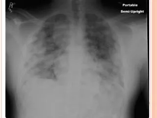
Diagnostic Testing • A complete history and physical can help to direct care. Initial evaluations should aim to locate source of bleeding and identify underlying cause. • Chest x-ray- Can help determine if patient has heart failure, collapsed lung, pneumonia, cystic fibrosis, emphysema, pulmonary edema, broken ribs, pneumothorax, congenital heart disease, problems with heart valves, or cancer (Mayo Clinic, 2011). • CT scan-useful in further investigating abnormalities on chest x-ray. Can detect abnormalities not seen on x-ray, as well as presence of tumors, excess fluid around lungs, pulmonary embolism, tuberculosis, COPD, bronchiectasis, pneumonia, congenital abnormalities, and interstitial lung disease (“Computed Tomography-Chest”, 2013).
Diagnostic Testing • Bronchoscopy is a procedure that allows the doctor to look inside the bronchi and bronchioles of the lungs. • Bronchoscopy can find tumors, signs of infection, excess mucus in airways, site of bleeding, and blockages. (National Heart, Lung, & Blood Institute [NHLBI], 2012).
Laboratory Studies • CBC, PT, INR, PTT, and ESR. • Gram’s stain of sputum. • Acid-fast stain • Sputum cytology • PPD • HIV testing (Uphold & Graham, 2003).
Management • Focuses on bleeding cessation, aspiration prevention, and treatment of underlying causes. • Airway, breathing, and circulation are top priority when evaluating patients.
Management • Non-massive hemoptysis-not emergent • Chest x-ray • CBC with differential • Sputum culture for acid-fast bacilli if TB is suspected. • Sputum C&S for pneumonia and lung abscess. • CT and MRI may detect any abnormalities unrecognized on chest x-ray. (Bidwell & Pachner, 2005).
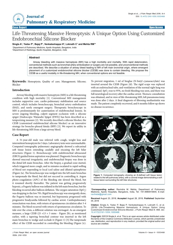
Management • Massive hemoptysis-over 600ml blood/24 hours. • Considered life threatening and is a medical emergency. • Requires immediate treatment, surgery, or bronchoscopy. • Management should focus on managing cardiorespiratory parameters, correction of hypoxia, stabilization of blood pressure, and blood transfusions if necessary. Surgical intervention • Bronchial artery embolization • Surgery-lobectomy or pneumonectomy
Pharmacological Management • Antibiotics for underlying bacterial or fungal infections. • Antitubercular drugs • Chemotherapy or radiation for lung cancer. • Steroids for inflammatory conditions.
Complications of hemoptysis • Asphyxia • Shock • Anemia • Renal failure • Atelectasis • Pulmonary infection
Follow-up/Referrals • Minimal hemoptysis related to respiratory infection should follow-up in 2-3 days if sputum is still blood-tinged (Uphold & Graham, 2003). • Blood-tinged sputum that is more than minimal will require a follow-up in 12-48 hours (Uphold & Graham, 2003). • Patients who present with non-massive hemoptysis, normal chest x-ray, and are considered low risk can be treated on an outpatient basis. • Pulmonology-recurrent or unexplained hemoptysis, COPD, cystic fibrosis. • Hematology/Oncology-diagnosed with malignancy or have coagulation disorder. • Cardiology-congestive heart failure, congenital heart defect, or mitral valve stenosis
Education • Patients and family should be educated regarding the causes of hemoptysis (Dunphy et al., 2011). • Notation of any change in color, amount, and consistency of blood expectorated should be reported to healthcare provider. Any increase in amount should be reported immediately (Dunphy et al., 2011). • Smoking cessation • If prescribed antibiotics, take all medication as directed by provider. • Pneumococcal vaccine • Proper hand washing to prevent spread of infection. • Referral to the American Lung Association website
Literature review • All the literature found regarding hemoptysis was very similar. Each article reviewed the disease processes that were associated with hemoptysis including evaluation, treating, and managing symptoms and disease. • There was no clear, concise number to differentiate between mild, moderate, and severe hemoptysis. Each author used their own definition, but all state that massive hemoptysis should be treated as a life-threatening condition.
Questions • Management of hemoptysis focuses on: a) Bleeding cessation b)Treatment of underlying cause c) Aspiration prevention d) All of the above 2) Underlying inflammatory disease of the tracheobronchial mucosa causes_____% of hemoptysis cases? a) 25% b) 60% c) 80% d) 12%
Questions 3) What is the difference between hemoptysis and hematemesis? a) Hemoptysis is the coughing up of purulent sputum, hematemesis is the coughing up of blood. b)Hemoptysis is the vomiting of blood, hematemesis occurs after you cut yourself while on anticoagulant therapy. c)Hemoptysis is the vomiting of blood, hematemesis is the coughing up of blood. d)Hemoptysis is the coughing up of blood, hematemesis is vomiting of blood. 4) Which is not a causative factor of hemoptysis? a) gastric ulcer b) Pneumonia c) Wegener granulomatosis d) Mycetoma
Questions 5) Epistaxis and expectoration of blood without a cough usually results from: a) Intestinal bleeding b) Lower respiratory source c) Upper respiratory source d) Gastric bleeding 6) Clinically, patients who present with undiagnosed lung abscess typically have which of the following signs and symptoms? a) Hemoptysis, fever, syncope b) Hemoptysis, fever, tachycardia, dyspnea on exertion c) Hemoptysis, ankle edema, JVD d) Hemoptysis, recurrent lower respiratory infections, copious amounts of purulent sputum
Questions 7) Which is the number one priority when a patient presents with a chief complaint of “coughing up blood?” a) Make sure that height and weight are taken b) Airway, breathing, and circulation are uncompromised c) Did they bring a sputum sample to visit? d) Vital signs 8) Which are the most important social/environmental aspects that you want to address when completing the patient’s history portion of exam? a) Sexual History b) Environmental exposure to asbestos c) Travel History d) All of the above
Questions 9) A patient presents to the ER with complaints of hemoptysis, sudden onset of pleuritic pain. Upon exam, you notice that patient is tachypneic, tachycardic, diaphoretic, cyanotic, fever, and 4th heart sound. What is your first assumption? a) Lung abscess b) Pulmonary infarction secondary to pulmonary emboli c) Pneumonia d) Chronic bronchitis 10) What are laboratory tests that can be ordered to diagnose cause of hemoptysis? a) Thyroid studies, lipid panel. b) Hepatic panel, BMP, HgA1C c) CMP, hCG level d) CBC, ESR, PT, PTT, INR
References Bidwell, J.L. & Pachner, R.W. (2005). Hemoptysis: Diagnosis and management. American Family Physician 72(7), 1253-1260. Retrieved from http://www.aafp.org/afp/2005/1001/p1253.html Computed tomography- chest (2013). Retrieved from October 6, 2013, from www.radiologyinfo.org/en/info.cfm?pg=chestct Corey, R. (2009). Hemoptysis. Clinical methods: The history, physical and laboratory examinations (3rd ed.). Retrieved from http://www.ncbi.nlm.nih.gov/books/NBK360/ Devine, S.T. & Lippmann, M. (2006). Management of massive hemoptysis. Respiratory Emergencies. Boca Raton, Fl: Taylor and Francis Group. Dunphy, L.M., Winland-Brown, J.E., Porter, B.O., & Thomas, D.J. (2011). Primary care: The art and science of advanced practice nursing (3rd ed.). Philadelphia, PA: F.A. Davis Company. Gaude, G.S. (2010). Hemoptysis in children. Indian Pediatrics 47(3), 245-254. Goroll, A.H., & Mulley, Jr., A.G. (2009). Primary care medicine: Office evaluation and management of the adult patient. Philadelphia, PA: Lippincott, Williams, & Wilkins. Mayo Clinic (2011). Chest x-rays. Retrieved from www.mayoclinic.com/health/chest-x-rays/MY00297
References National Heart, Lung, & Blood Institute (2012). What is bronchoscopy? Retrieved from www.nhlbi.nih.gov/health/health-topics/topics/bron/ National Library of Medicine (2011). Goodpasture syndrome. Retrieved from http://www.nlm.nih.gov/medlineplus/ency/article/000142.htm Uphold, C.R. & Graham, M.V. (2003). Clinical guidelines in family practice (4th ed.). Gainesville, FL: Barmarrae Books, Inc. Wise, C.M. (2013). Wegener granulomatosis. In F.J. Domino (21st ed.). The 5-minute clinical consult 2013. Philadelphia, PA: Lippincott, Williams, & Wilkins. Whitehurst-Cook, M. (2013). Gastritis. In F.J. Domino (21st ed.). The 5-minute clinical consult 2013. Philadelphia, PA: Lippincott, Williams, & Wilkins.