Download
1 / 24
250 likes | 914 Vues
Case Presentation. The patient is a 30 year-old Pakistani woman with CF who presents complaining of large-volume hemoptysis.The patient was relatively well until 1 month PTA when she developed an increasing productive cough. Her FEV1 was noted to be 1.4 (pt's baseline: 1.9). Her sputum grew Pseudom

E N D
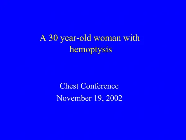
1. A 30 year-old woman with hemoptysis Chest Conference
November 19, 2002
2. Case Presentation The patient is a 30 year-old Pakistani woman with CF who presents complaining of large-volume hemoptysis.
The patient was relatively well until 1 month PTA when she developed an increasing productive cough. Her FEV1 was noted to be 1.4 (pt�s baseline: 1.9). Her sputum grew Pseudomonas and she was treated for 2 weeks with IV Tobra/Timentin. She had scant hemoptysis during this episode that resolved with antibiotics.
She was off antibiotics and without pulmonary symptoms until 1 day PTA when she awoke choking and coughing up large amounts of blood. She states she coughed up about 200cc of blood over 15-20 minutes, then blood streaked sputum for several hours.
3. Case Presentation On presentation to the ED, she endorsed orthostatic symptoms, but denied fever, chills, recent productive cough, SOB and chest pain.
4. Case Presentation Past Medical History
Cystic Fibrosis: sweat test neg; F508 mutation +; + colonization with drug-resist Pseudomonas. Admitted for hemoptysis in 1998; resolved w/ medical treatment.
Iron deficiency anemia
Medications
Salmeterol
Ipratropium/albuterol
Fluticasone
Social History: lives in Morgan Hill, CA with partner; no drugs, EtOH or tobacco
5. Case Presentation Physical Examination
36.7 110/71 95 15 99% RA
Gen: well-appearing woman in NAD
HEENT: no oropharyngeal lesions; no LAD
Lungs: few rales @ left mid-lung
CV: no R/M/G
Abdomen: soft, NT, ND
Ext no edema
6. Case Presentation Labs:
WBC: 14
Hct: 35 (baseline Hct: 35)
Platelets: 332
Coags: normal
EKG: sinus rhythm @ 85
9. Hospital Course Pt was admitted; treated with IV tobramycin & Timentin
Scant hemoptysis x 8 hours in the ED
Hct: 35 --> 31.
10. Hospital Course Pt underwent selective bronchial arteriography :
Aortogram demonstrates two large bronchial arteries. One bronchial artery supplies the mid and upper right lung. The other bronchial artery supplies the left mid and upper lung. These two vessels appear to have a common trunk origin.
The bronchial arteries were successfully embolized with polyvinyl alcohol particles (350-500 and 500-710 microns).
Follow-up angiographic run demonstrates some persistent flow of the right bronchial artery to the region along the medial apex.
11. Hospital Course The patient developed severe R pleuritic CP, temp to 39.0C and mild dysphagia.
Temperature and dysphagia resolved within 5 hours and chest wall pain resolved with low doses of opiates.
The patient had no further episodes of hemoptysis and was discharged on HD#3 with a 14 day course of IV antibiotics.
13. Bronchial Artery Embolization in CF
14. Bronchial Artery Hypertrophy Figure 1.�Bronchial artery hypertrophy
A, Bronchial arteriogram of a patient with hemoptysis from diffuse bronchiectasis. Note the extensive dilation, hypertrophy, and proliferation of the bronchial arteries (closed arrows) whose diameters are normally no larger than those of the posterior intercostal arteries (open arrows). B, A close-up view of right lung bronchial arteriogram. Same patient as in A, following selective bronchial artery embolization.Figure 1.�Bronchial artery hypertrophy
A, Bronchial arteriogram of a patient with hemoptysis from diffuse bronchiectasis. Note the extensive dilation, hypertrophy, and proliferation of the bronchial arteries (closed arrows) whose diameters are normally no larger than those of the posterior intercostal arteries (open arrows). B, A close-up view of right lung bronchial arteriogram. Same patient as in A, following selective bronchial artery embolization.
15. Marshall et al. Eur Rad. 1997;7:1221-7 Bronchial Artery Anatomy Significant variability in origin of bronchial arteries.
70% arise from the thoracic aorta at T5-6.
45% have 2 right BA and a single left BA.
Up to 15% have anomalous BA origins (thyrocervical trunk, internal mammary, inferior phrenic).
2-50% have a spinal artery branching off a bronchial artery.
Bronchial arteries supply: bronchi, middle third of the esophagus, diaphragmatic and visceral mediastinal pleura, aortic vasa vasorum, and spinal cord.
16. Brinson et al. AJRCCM. 1998; 157:1951-8 Complications of Bronchial Artery Embolization Minor complications occur in 10-30% (fever, chest wall pain, dysphagia)
Major complications in various case reports:
Bronchial necrosis
Bowel ischemia
Transverse myelitis (transient)
Paraplegia related to spinal artery embolization (less of a concern with microcatheter technology).
17. Spinal Artery Embolization
18. Stern et al. Am Rev Resp Dis. 1978; 117:825-28 Medical Management of Massive Hemoptysis in Cystic Fibrosis 728 patients with CF at an academic center
38/728 (5%) had massive hemoptysis (>300ml/24hrs)
All pts treated w/ antibiotics, vit K and fluids
5 pts required transfusions
4 had transient hypotension
Hemoptysis stopped in all subjects without surgical intervention
34/38 had resolution of hemoptysis within 96hrs
10/38 died during follow-up (mean survival 2.5 years); survival rate was similar to that of control group without massive hemoptysis.
17/38 had recurrent massive hemoptysis in follow-up.
19. Stern et al. Am Rev Resp Dis. 1978;117:825-28 Medical Management of Massive Hemoptysis in Cystic Fibrosis Conclusion:
�Because patients with massive hemoptysis tend to stop bleeding without surgical intervention, future studies of therapies for massive hemoptysis must have appropriate control patients who receive only medical management.�
20. Cohen et al. Radiology; 1990;175:401-05 BAE to Control Hemoptysis in CF 20/425 CF patients developed hemoptysis from 1982-86.
Indications for BAE: failure of med management and
1. An episode of massive hemoptysis w/ persistent bleeding.
2. 3 or more 100mL hemorrhages within 1 week.
3. Chronic hemoptysis interfering with lifestyle.
4. Hemoptysis preventing effective postural drainage.
21. Cohen et al. Radiology. 1990;175:401-05 BAE to Control Hemoptysis in CF Results: 85% (19/20) had immediate control of bleeding with BAE
35% (7/20) had aberrant bronchial arteries.
55% (11/20) had a spinal artery coming off a bronchial artery.
90% 17/19 developed post-procedure chest pain, fever, or dysphagia.
No assessment of long term rate of re-bleed.
No matched control group.
22. Sweezey et al. Chest. 1990; 97:1322-26. BAE for Massive Hemoptysis in CF Case �control study of 25 pts with CF and massive hemoptysis.
Control group: hemoptysis-free; matched for disease severity, age and sex.
84% (21/25) had immediate control of hemoptysis with BAE.
6 died within 3 months of embolization.
Reduced survival in embolized group vs control (p<0.02)
52% percent had recurrent massive hemoptysis (mean 20.5 months after first BAE).
52% had chest wall pain and fever after BAE. No serious complications.
23. Sweezey et al. Chest. 1990:97:1322-26 BAE for Massive Hemoptysis in CF Conclusions:
BAE effective in controlling massive hemoptysis.
BAE has a low risk of serious adverse effects.
BAE does not improve mortality, but may increase time to recurrent massive hemoptysis.
24. Schidlow et al. Ped Pulmon. 1993;15:187-98 Cystic Fibrosis Foundation Treatment Guidelines for Major Hemoptysis �There are almost no scientific data to support most of the therapeutic options listed for treatment of hemoptysis in CF.�
Discontinuation of drugs which could interfere with anticoagulation and reversal of coagulation abnormalities.
Transfusions as clinically indicated.
Treatment with appropriate antibiotics based on recent sputum cultures.
Arterial embolization may be indicated for major hemoptysis or for minor hemoptysis when it interferes with patient�s lifestyle or medical management.