Nomenclature
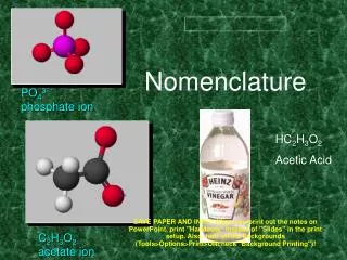
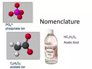
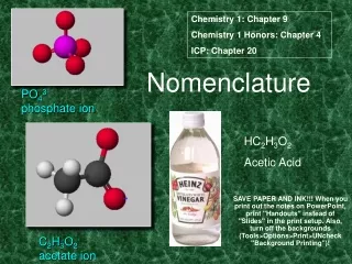
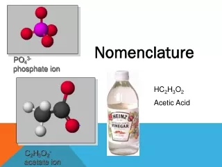
Nomenclature. Nucleoside Nucleotide Base + deoxyribose +phosphate. Purines adenine adenosine guanine guanosine hypoxanthine inosine Pyrimidines thymine thymidine cytosine cytidine +ribose uracil uridine. ii). Structure of the DNA double helix.
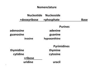

Nomenclature
E N D
Presentation Transcript
Nomenclature Nucleoside Nucleotide Base +deoxyribose +phosphate Purines adenine adenosine guanine guanosine hypoxanthine inosine Pyrimidines thymine thymidine cytosine cytidine +ribose uraciluridine
ii). Structure of the DNA double helix Structure of the DNA polynucleotide chain 5’ 3’ • polynucleotide chain • 3’,5’-phosphodiester bond
ii). Structure of the DNA double helix • The DNA double helix requires that the two polynucleotide chains be base-paired to each other.
X ray diffraction • When X rays are focused through isolated macromolecules or crystals of purified molecules, the X ray are deflected by the atom of the molecules in specific patterns called diffraction patterns. • It provides the information about the organization of the components of the molecules. • Watson and Crick had X ray crystallographic data on DNA structure from the studies of Wilkins and Franklin and their coworkers. • These data indicated that DNA was a highly ordered, multiple stranded structure with repeating sub structures spaced every 3.4 Ao (1 Angstrom = 10-10 m )
X-ray diffraction patterns of DNA – Rosalind Franklin and Maurice Wilkins The central cross shaped pattern as indicative of a helical structure. The heavy dark patterns (top and bottom) indicate that the bases are stacked perpendicular to the axis of the molecule.
A-T base pair Hydrogen bonding of the bases G-C base pair Chargaff’s rule: The content of A equals the content of T, and the content of G equals the content of C in double-stranded DNA from any species
Double-stranded DNA • Double-stranded DNA, is composed of two base-paired, complementary polynucleotide chains. • Base-pairing between the complementary strands is required for two important functions of DNA: 1) DNA replication involves an unwinding of the double helix (right) followed by synthesis of a complementary strand from each of the unpaired template strands, and 2) RNA synthesis DNA serves as a template for by utilizing the information in one strand to code for a complementary RNA strand.
DNA in the "B" form has - a major groove and a minor groove, -and has 10 base pairs per one turn of the double helix. • “Supercoiled" DNA that is overwound or underwound, with fewer than or more than 10 base pairs per turn. • Antiparallel with respect to each other. • Each polynucleotide chain has a 5' end and a 3' end.
Double-stranded DNA 5’ 3’ Major groove Minor groove “B” DNA 3’ 5’ 3’ 5’
Deoxyribonucleases (or DNases) are enzymes that cleave phosphodiester bonds. • Some are used for constructive purposes, such as proofreading during DNA replication, whereas others are used to degrade DNA. • There are two basic classes of DNases: exonucleases and endonucleases. Exonucleases remove only the terminal nucleotide, whereas endonucleases cleave anywhere within the DNA double helix.
Forces affecting the stability of the DNA double helix • hydrophobic interactions - stabilize • - hydrophobic inside and hydrophilic outside • stacking interactions - stabilize • - relatively weak but additive van der Waals forces • hydrogen bonding - stabilize • - relatively weak but additive and facilitates stacking • electrostatic interactions - destabilize • - contributed primarily by the (negative) phosphates • - affect intrastrand and interstrand interactions • - repulsion can be neutralized with positive charges • (e.g., positively charged Na+ ions or proteins)
Stacking interactions Charge repulsion Charge repulsion
Denaturation of DNA Strand separation and formation of single-stranded random coils Double-stranded DNA Extremes in pH or high temperature A-T rich regions denature first Cooperative unwinding of the DNA strands
Electron micrograph of partially melted DNA Double-stranded, G-C rich DNA has not yet melted A-T rich region of DNA has melted into a single-stranded bubble • A-T rich regions melt first, followed by G-C rich regions
Hyperchromicity Absorbance maximum for single-stranded DNA Absorbance maximum for double-stranded DNA Absorbance 220 260 300 The absorbance at 260 nm of a DNA solution increases when the double helix is melted into single strands.