Download
1 / 91
910 likes | 1.15k Vues
BIOLOGICAL MEMBRANES AND PRINCIPLES OF SOLUTE AND WATER MOVEMENT. Carmel M. McNicholas, Ph.D. Department of Physiology & Biophysics Contact Information: MCLM 868 934 1785 cbevense@uab.edu. Sept. ‘11. OUTLINE Biological Membranes and Principles of Solute and Water Movement

E N D
BIOLOGICAL MEMBRANES AND PRINCIPLES OF SOLUTE AND WATER MOVEMENT Carmel M. McNicholas, Ph.D.Department of Physiology & BiophysicsContact Information:MCLM 868934 1785cbevense@uab.edu Sept. ‘11
OUTLINE • Biological Membranes and Principles of Solute and Water Movement • Diffusion and Osmosis • Principles of Ion Movement • Membrane Transport • Nerve Action Potential • HANDOUT AND PROBLEM SET
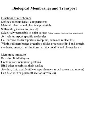
The Cell: The basic unit of life (i) obtaining food and oxygen, which are used to generate energy (ii) eliminating waste substances (iii) protein synthesis (iv) responding to environmental changes (v) controlling exchange of substances (vi) trafficking materials (vii) reproduction.
The fluid compartments of a 70kg adult human EXTRACELLULAR (~40%) INTRACELLULAR (~60%) INTERSTITIAL FLUID ~13 L BLOOD PLASMA ~3 L [Na+] = 145 mM [K+] = 4.5 mM [Cl-] = 116 mM [protein] = 0 mM Osmolality = 290 mOsm INTRACELLULAR FLUID ~25 L [Na+] = 142 mM [K+] = 4.4 mM [Cl-] = 102 mM [protein] = 1 mM Osmolality = 290 mOsm [Na+] = 15 mM [K+] = 120 mM [Cl-] = 20 mM [protein] = 4 mM Osmolality = 290 mOsm TRANSCELLULAR FLUID ~1 L Composition: variable Epithelial cells Capillary endothelium Plasma membrane TOTAL BODY WATER (~42 L) Modified from: Boron & Boulpaep, Medical Physiology, Saunders, 2003.
Solute composition of key fluid compartments • Osmolality constant • Cell proteins – 10-20% of the cell mass • Structural and functional
Membranes are selectively permeable Gas molecules are freely permeable Gas molecules are freely permeable Gas molecules are freely permeable Gas molecules are freely permeable Gas molecules are freely permeable Small uncharged molecules are freely permeable Small uncharged molecules are freely permeable Small uncharged molecules are freely permeable Large / charged molecules need ‘assistance’ to traverse the plasma membrane Large / charged molecules need ‘assistance’ to traverse the plasma membrane Large / charged molecules need ‘assistance’ to traverse the plasma membrane
The Extracellular Matrix Epithelial cell The extracellular matrix (ECM) of animal cells functions in support, adhesion, movement and regulation Basement membrane Capillary endothelium Fibroblast Connective tissue and ECM
The Extracellular Matrix • The ECM is an organized meshwork of polysaccharides and proteins secreted by fibroblasts. Commonly referred to as connective tissue. • COMPOSITION: • Proteins: Collagen (major protein comprising the ECM), fibronectin, laminin, elastin • Two functions: structural or adhesive • Polysaccharides: Glycosaminoglycans, which are mostly found covalently bound to protein backbone (proteoglycans). • Cells attach to the ECM by means of transmembrane glycoproteins called integrins • Extracellular portion of integrins binds to collagen, laminin and fibronectin. • Intracellular portion binds to actin filaments of the cytoskeleton
The Cytoskeleton Intracellular network of protein filaments • Role • Supports and stiffens the cell • Provides anchorage for proteins • Contributes to dynamic whole cell activities (e.g., dividing and crawling of cells and moving vesicles and chromosomes) • Three Types Of Cytoskeletal Fibers • Microtubules (tubulin - green) • Microfilaments (actin-red) • Intermediate filaments
Structural Junctions Tight Junctions Adhering Junctions Desmosome Zonula Adherens (belt)
Gap Junctions • ROLE: Passage of solutes (MW<1000) from cell to cell. • Cell-cell communication • Propagation of electrical signal
The Membrane Glycocalyx - cell coat Alberts et al., Molecular Biology of the Cell, 4th Ed. Garland Science, 2002) • Carbohydrates are: • Covalently attached to membrane proteins and lipids • Sugar chains added in the ER and modified in the golgi • Oligo and polysaccharide chains absorb water and form a slimy surface coating, which protects cell from mechanical and chemical damage. • Membrane Carbohydrates and Cell-Cell Recognition • crucial in the functioning of an organism. It is the basis for: • Sorting embryonic cells into tissues and organs. • Rejecting foreign cells by the immune system.
Transport of large molecules EXOCYTOSIS: Transport molecules migrate to the plasma membrane, fuse with it, and release their contents. ENDOCYTOSIS:The incorporation of materials from outside the cell by the formation of vesicles in the plasma membrane. The vesicles surround the material so the cell can engulf it.
Membranes are selectively permeable Gas molecules are freely permeable Gas molecules are freely permeable Gas molecules are freely permeable Gas molecules are freely permeable Gas molecules are freely permeable Small uncharged molecules are freely permeable Small uncharged molecules are freely permeable Small uncharged molecules are freely permeable Large / charged molecules need ‘assistance’ to traverse the plasma membrane Large / charged molecules need ‘assistance’ to traverse the plasma membrane Large / charged molecules need ‘assistance’ to traverse the plasma membrane
Diffusion Diffusion is the net movement of a substance (liquid or gas) from an area of higher conc. to one of lower conc. due to random thermal motion.
Kinetic characteristic of diffusion of an uncharged solute Model: compartments separated by permeable glass x Cs1 Cs2 compartment 1 compartment 2 A = cross sectional area of the glass disc Cs = concentration of uncharged solute x = thickness
x Cs1 Cs2 compartment 1 compartment 2 According to kinetics, the rate of movement can be described as follows: rate of diffusion from 1 2 = kCs1 -{rate of diffusion from 2 1 = kCs2} ---------------------------------------------------------------------------- net rate of diffusion across barrier = k(Cs1-Cs2) = kCs where k is a proportionality constant.
Diffusion is proportional to the surface area of the barrier (A) and inversely proportional to its thickness (x). k can thus be expressed as ADs/x, where Ds is the diffusion coefficient of the solute. The concentration gradient across the membrane is the driving force for net diffusion.
FLUX (Js) describes how fast a solute moves, i.e. the number of moles crossing a unit area of membrane per unit time (moles/cm2.s) Therefore, net diffusion rate = ADsCs/x. Dividing both sides by A (to obtain flux), we obtain: Fick’s first law of diffusion: Flux = Js = DsCs/x “The rate of flow of an uncharged solute due to diffusion is directly proportional to the rate of change of concentration with distance in direction of flow” When the concentration gradient of a substance is zero the system must be in equilibrium and the net flux must also be zero.
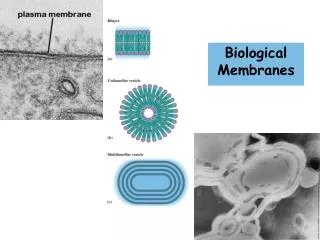
Diffusion of an uncharged soluteModel: compartments separated by a lipid bilayer x Cs1 Cs2 compartment 1 compartment 2 Biological membranes are composed of a lipid bilayer of phospholipids interspersed with integral and peripheral proteins (“Fluid Mosaic Model”).
Partitioning of an uncharged solute across a lipid bilayer The partition coefficient, Ks will increase or decrease the driving force of the solute S across the membrane: Js = KsDsCs/x Because it is difficult to measure Ks, Ds and x, these terms are often combined into a permeability coefficient, Ps = KsDs/x. It follows that: Js = PsCs Cs1 Lipophilic Ks > 1 Ks lies between 0 and 1 Cs2 Hydrophilic Ks < 1
Solute movement across a lipid bilayer through entry into the lipid phase occurs by simple diffusion. This movement occurs downhill and is passive.
Osmosis: The flow of volume Osmosis refers to the net movement of water across a semi-permeable membrane (or displacement of volume) due to the solute concentration difference.
Osmosis. The flow of volume The solute concentration difference causes water to move from compartment 2 1. The pressure required to prevent this movement is the osmotic pressure. Time 1 2 1 2
Osmosis. The flow of volume AN IDEAL MEMBRANE (Meniscus) Piston H2O Cs1 Cs2 (Compartment 2 is open to the atmosphere) (The piston applies pressure to stop water flow) Compartment 1 Compartment 2 Here the membrane is only permeable to water which will flow down its concentration gradient from 2 1. The volume flow can be prevented by applying pressure to the piston. The pressure required to stop the flow of water is the osmotic pressure of solution 1.
The osmotic pressure () required is determined from the van’t Hoff equation: = RTCS = (25.4)CSatm at 37°C. Where, R = the gas constant (0.082 L.atm.K-1.mol-1), T = absolute temperature (310 K @ 37 ºC) and CS (mol.L-1) is the concentration difference of the uncharged solute
Osmosis. Importance of osmolarity φic = osmotically effective concentration φ is the osmotic coefficient ‘i’ is the number of ions formed by dissociation of a single solute molecule ‘c’ is the molar concentration of solute (moles of solute per liter of solution) e.g. what is the osmolarity of a 154 mM NaCl solution, where φ = 0.93 → 154 x 2 x 0.93 = 286.4 mOsm/l
Osmosis. The flow of volume A NONIDEAL MEMBRANE H2O Piston S Cs1 Cs2 • The osmotic pressure depends on the ability of the membrane to distinguish between solute and solvent. • If the membrane is entirely permeable to both, then intercompartmental mixing occurs and = 0. • The ability of the membrane to “reflect” solute S is defined by a reflection coefficient Sthat has values from 0 (no reflection) to 1 (complete reflection). • Thus, the effective osmotic pressure for nonideal membranes is: • eff = SRTCS
Osmotic and hydrostatic pressure differences in volume flow Volume flow across a membrane is described by: JV = KfP where Kf is the membrane’s hydraulic conductivity and P is the sum of pressure differences. These pressure differences can be hydrostatic (PH), osmotic (eff) or a combination of both. There is equivalence of osmotic and hydrostatic pressure as driving forces for volume flow, hence Kf applies to both forces. Thus, JV = Kf(eff – PH)(Starling equation) and (eff – PH) is the driving force for volume flow.
Starling Forces Venule Arteriole Interstitial space = fluid movement Interstitial fluid pressure under normal conditions ~0 mmHg Hydrostatic pressure Filtration dominates Absorption dominates Osmotic (oncotic) pressure Importance of plasma proteins!
Diffusion of Electrolytes K+ Ac- Cs1=100mM Cs2=10mM V – + For charged species, both electrical and chemical forces govern diffusion.
The Principle of Bulk Electroneutrality All solutions must obey the principle of bulk electroneutrality: the number of positive charges in a solution must be the same as the number of negative charges.
Diffusion of Electrolytes K+ Cs1=100mM Cs2=10mM Ac- Ac- K+ V – + Law of electroneutrality (for a bulk solution) must be maintained. In the above model in which the membrane becomes permeable to sodium (K+) and acetate (Ac–), both ions will move from side 1 2. The concentration gradient between compartment 1 and 2 is the driving force. K+ (with the smaller radius) will move slightly ahead of Ac–, thereby creating a diffusing dipole. A series of dipoles will generate a diffusion potential. Eventually, equilibrium is reached and Cs1 = Cs2 = 55mM
Diffusion of Electrolytes K+ Cs1=100mM Cs2=10mM Ac- V – + When the membrane is permeable to only one of the ions (e.g., K+) an equilibrium potential is reached. Here, the chemical and electrical driving forces are equal and opposite. Equilibrium potentials (in mV) are calculated using the Nernst equation: R = gas constant; T = absolute temp.; F = Faraday’s constant; z = charge on the ion (valence); 2.3RT/F = 60 mV at 37ºC
The Nernst Equation is satisfied for ions at equilibrium and is used to compute the electrical force that is equal and opposite to the concentration force. At the Nernst equilibrium potential for an ion, there is no net movement because the electrical and chemical driving forces are equal and opposite. • Even when there is a potential difference across a membrane, charge balance of the bulk solution is maintained. • This is because potential differences are created by the separation of a few charges adjacent to the membrane.
Calculating a Nernst Equilibrium Potential Na+ Cs1 = 100mM Cs2 = 10mM Ac- V – + For the model above, the Nernst potential for Na+, ENa = 60 log(100/10) = +60 mV
Taking valence of the ion into account in calculating a Nernst potential [Cl-]i = 10 mM Here, z = -1 [Cl-]o = 100 mM
[K+]i = 100 mM [K+]o = 10 mM Equilibrium potentials of various ions for a mammalian cell
Remember: Log 10/100 = log 0.1 = –1 Log 100/10 = log 10 = +1 A 10-fold concentration gradient of a monovalent ion is equivalent, as a driving force, to an electrical potential of 60 mV.
Membrane potential vs. equilibrium potential When a cell is permeable to more than one ion then all permeable ions contribute to the membrane potential (Vm).
1. Most biologic membranes are virtually impermeable to: • Hydrophilic molecues having molecular radii > 4Å e.g. glucose, amino acids) • Charged molecules • 2. The intracellular concentration of many water soluble solutes differ from the medium in which they are bathed. • Thus, mechanisms other than simple diffusion across the lipid bilayer are required for the passage of solutes across the membrane.