Precipitation Tests
Precipitation Tests. Lattice Formation. Ab in gel. Ag. Ag. Ag. Ag. Diameter 2. Ag Concentration. Radial Immunodiffusion (Mancini). Method Ab in gel Ag in a well. Interpretation Diameter of ring is proportional to the concentration Quantitative Ig levels. -. +. Ag. Ag. Ab.


Precipitation Tests
E N D
Presentation Transcript
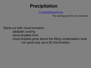
Precipitation Tests Lattice Formation
Ab in gel Ag Ag Ag Ag Diameter2 Ag Concentration Radial Immunodiffusion (Mancini) • Method • Ab in gel • Ag in a well • Interpretation • Diameter of ring is proportional to the concentration • Quantitative • Ig levels
- + Ag Ag Ab Ag Ab Immunoelectrophoresis • Ab is placed in trough cut in the agar • Method • Ags are separated by electrophoresis • Interpretation • Precipitin arc represent individual antigens
Immunoelectrophoresis • Method • Interpretation • Qualitative • Relative concentration
- + Ab Ag Countercurrent electrophoresis • Method • Ag and Ab migrate toward each other by electrophoresis • Used only when Ag and Ab have opposite charges • Qualitative • Rapid
Radioimmuoassays (RIA)Enzyme-Linked Immunosorbent Assays (ELISA) Lattice formation not required
Prior to Test Y Y + Labeled Ag Test Y Y + + + Labeled Ag Patient’s sample Competitive RIA/ELISA for Ag • Method • Determine amount of Ab needed to bind to a known amount of labeled Ag • Use predetermined amounts of labeled Ag and Ab and add a sample containing unlabeled Ag as a competitor
Solid Phase Solid Phase Test Y Y + + + Labeled Ag Patient’s sample Competitive RIA/ELISA for Ag • Method cont. • Determine amount of labeled Ag bound to Ab • NH4SO4 • anti-Ig • Immobilize the Ab • Concentration determined from a standard curve using known amounts of unlabeled Ag • Quantitative • Most sensitive test
Labeled Anti-Ig Ab in Patient’s sample Y Y Ag Immobilized Solid Phase Solid Phase Non-Competitive RIA/ELISA • Ab detection • Immobilize Ag • Incubate with sample • Add labeled anti-Ig • Amount of labeled Ab bound is proportional to amount of Ab in the sample • Quantitative
Labeled Ab Ag in Patient’s sample Y Ag Y Immobilized Solid Phase Solid Phase Non-Competitive RIA/ELISA • Ag detection • Immobilize Ab • Incubate with sample • Add labeled antibody • Amount of labeled Ab bound is proportional to the amount of Ag in the sample • Quantitative
Tests for Cell Associated Antigens Lattice formation not required
Fluorochrome Labeled Ab Y Ag Tissue Section Immunofluorescence • Direct • Ab to tissue Ag is labeled with fluorochrome
Fluorochrome Labeled Anti-Ig Unlabeled Ab Y Y Ag Tissue Section Immunofluorescence • Indirect • Ab to tissue Ag is unlabeled • Fluorochrome-labeled anti-Ig is used to detect binding of the first Ab. • Qualitative to Semi-Quantitative
Flow Tip FL Detector Light Scatter Detector Laser Immunofluorescence • Flow Cytometry • Cells in suspension are labeld with fluorescent tag • Direct or Indirect Fluorescence • Cells analyzed on a flow cytometer
Two Parameter Histogram Green Fluorescence Intensity Red Fluorescence Intensity Immunofluorescence • Flow Cytometry cont. • Data displayed One Parameter Histogram Unstained cells FITC-labeled cells Number of Cells Green Fluorescence Intensity
Assays Based on Complement Lattice formation not required
No Ag Ag Patient’s serum Y Y Y Y Y Y Y Y Y Y Complement Fixation • Methodology • Standard amount of complement is added • Ag mixed with test serum to be assayed for Ab • Erythrocytes coated with Abs is added • Amount of erythrocyte lysis is determined Ag Ag