Download
1 / 24
280 likes | 370 Vues
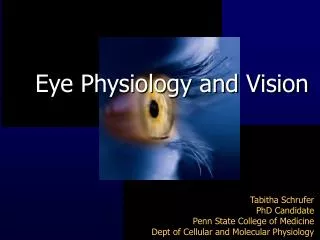
Dive into the intricate anatomy of the eye, explore retinal functions, neural pathways, and optical principles to grasp the complexity of vision physiology. Learn about visual receptors, neural pathways, and the principles of optics.

E N D
Anatomy of the eye • 1- sclera: is the outer protective layer. • 2- cornea : anterior , modified part of the sclera, light rays enter through it. • 3- choroid : deep to the sclera , rich in blood vessels. • 4-retina : lines the post. Two thirds of the choroid , formed of neural tissue rich in receptors. • 5- cilliary bodies : thickened anterior parts of the choroid , contain circular & longitudinal muscle fibers , produce aqueos humor. • 6-iris: pigmented , opaque , contains constrictors of the pupil ( circular ) & dilators of the pupil ( radial ).
Anatomy of the eye • 7- vitreous humor : a gelatinous substance between the lens & retina. • 8- aqueous humor : a clear liquid that nourishes the cornea & lens. • 9- canal of Schlemm : at the junction between the iris & cornea , drains aqueous humor.
Retina • Extends anteriorly almost reaching the cilliary body. • Contains visual receptors ( rods & cones) + bipolar , ganglion , horizontal & amacrine cells. • Rods & cones are next to the choroid . They synapse with bipolar cells . • Bipolar cells synapse with ganglion cells. • Amacrine cells connect the ganglion cells to each other. • Axons of the ganglion cells converge to form the optic nerve. • Optic disk : is the point where the optic nerve leaves the eye & blood vessels enter.It is located 3mm medial to & slightly above the posterior pole of the globe.
Retina • The optic disk is a blind spot , it doesn’t contain any visual receptors. • Macula lutea : • is a yellowish area near the posterior pole. It marks the location of the fovea centralis. • Fovea centralis : • a thin , rod-free , cone-packed area. • Each cone synapses with a single bipolar cell , which in turn synapses with a single ganglion cell. • Contains NO blood vessels. • Maximum visual acuity.
Retinal blood supply • Retinal vessels : supply the bipolar & ganglion cells. • Choroid plexus : supplies the rods & cones.
Neural pathways. • Axons of ganglion cells pass caudally , converge & form the optic nerve. • The optic nerve passes to the optic tract & ultimately reaching the lateral genicualte body of the thalamus. • Fibers from each nasal (medial) hemiretina decussate in the optic chiasm . • Fibers from the temporal ( lateral) hemiretina do not decussate. • In the geniculate body , fibers from one nasal hemiretina synapse with the temporal fibers of the other retina to form the geniculocalacrine tract. • The geniculoclacrine tract passes to the primary visual receiving area of the occipital lobe ( broadman’s 17 )
Receptors • Each rod & cone is divided into : 1- an outer segment : -made of modified cillia that form saccules & disks . • The saccules & disks contain photosensitive compounds. 2- an inner segment : -contains a nuclear region. -rich in mitochondria. 3- a synaptic zone.
Receptors • Rods have a thin , rod-like ( 27lf !) appearance in the outer segment. • Cones have thick inner segments & conical outer segments ( 27lf marrathanya! ) • Rods predominate in the extrfoveal portions of the retina.
Duplicity theory • Rods : - are extremely sensitive to light ( low threshold of stimulation ) • Are scotopic ( scoto= no light ) : they are used in night vision. • Are incapable of resolving details or colors. • Cones : • - have a much higher threshold , & much greater acuity. • Are photopic ( photo = light ) : used for light & color vision.
Principles of optics. • Light rays are refracted when passing from one medium to another due to the difference in density. • Parallel light rays striking a biconcave lens are refracted to a point behind the lens , called the principal focus.
Principles of optics • Principle focal distance : is the distance between the lens & principle focus. • Biconcave lenses diverge light , biconvex ones converge it. • Diopters : are used to measure the refractive power of a lens in meters • They are the reciprocal of the principle focal distance in meters. • 1 Diopter = 1/ PFL • The human eye has a refractive power of 6 m at rest.
Principles of optics • Diopteric power of the eye : • Cornea = 40-45 D • Lens = 15-20 D • Accommodation +14 ( depending on age )
Accommodation. • Is an active process that requires muscular actions. • At rest , objects closer than 6 m to the eye appear blurred. • This blurriness is diminished by accommodation. • So accommodation is : the process by which the curvature of the lens is increased in order to focus on a near object. • When looking at a near object , the cilliary muscles contract , the distance between the edges of the cilliary bodies decrease & the lens ligaments relax . Thus , the lens becomes more convex.
Accommodation When the cilary muscles are relaxed, the zonalus pulls tight and keeps the lens flattened for distant vision The elastic lens is attached to the circular cilary muscles by the zonalus which is made of inelastic fibres When the cilary muscles contract, it releases the tension on the zonalus and the elastic lens returns to a more rounded shape suitable for near vision
Near point • Is the nearest point to the eye where an object can be brought into clear focus by accommodation. • Recedes throughout life due to the gradual hardening of the lens. • This leads to presbyopia ( loss of lens elasticity )
Near response • Is a three-part response that consists of : 1- accommodation. 2- convergence of the visual axes. 3- pupillary constriction.
Pupillary reflexes. • Pupillary light reflex : When light is directed into the eye, its pupil constricts. • Consensual light reflex : When light is directed into one eye, the pupil of the other also constricts due to complicated mechanisms. • Pathway : Retina ---- optic tract ---- superior colliculus ---- occulomotor nucleus ---- pupillary muscles.
Argyll – Robertson pupils • Seen in some pathologies such as neurosyphyllis . • Characterized by loss of light reflex but not accommodation reflex.