Download
1 / 71
790 likes | 1.21k Vues
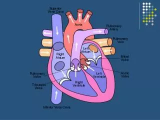
Physiology of the Heart. Review. The heart is a four-chambered muscular pump The body has two circulations; Pulmonary to the lungs and systemic to the entire body The atria receive blood from both circulations The ventricles send blood through the two circulations

E N D
Review The heart is a four-chambered muscular pump The body has two circulations; Pulmonary to the lungs and systemic to the entire body The atria receive blood from both circulations The ventricles send blood through the two circulations In order for the heart to pump it's blood, the heart must “beat” with rhythm
Cardiac Muscle Contraction Depolarization of the heart is rhythmic and spontaneous About 1% of cardiac cells have automaticity— (are self-excitable) Gap junctions ensure the heart contracts as a unit Long absolute refractory period (250 ms)
Cardiac Muscle Contraction Depolarization opens voltage-gated fast Na+ channels in the sarcolemma Reversal of membrane potential from –90 mV to +30 mV Depolarization wave in T tubules causes the SR to release Ca2+ Depolarization wave also opens slow Ca2+ channels in the sarcolemma Ca2+ surge prolongs the depolarization phase (plateau)
1 Action potential Depolarizationis due to Na+influx through fast voltage-gated Na+ channels. A positive feedback cycle rapidly opens many Na+ channels, reversing the membrane potential. Channel inactivation ends this phase. Plateau 2 Membrane potential (mV) Tension development (contraction) Tension (g) 3 1 2 Plateau phaseis due to Ca2+ influx through slow Ca2+channels. This keeps the cell depolarized because few K+channels are open. Absolute refractory period 3 Repolarizationis due to Ca2+channels inactivating and K+ channels opening. This allows K+efflux, which brings the membrane potential back to its resting voltage. Time (ms) Figure 18.12
Cardiac Muscle Contraction Ca2+ influx triggers opening of Ca2+-sensitive channels in the SR, which liberates bursts of Ca2+ Excitation-Contraction coupling occurs as Ca2+ binds to troponin and sliding of the filaments begins Duration of the AP and the contractile phase is much greater in cardiac muscle than in skeletal muscle Repolarization results from inactivation of Ca2+ channels and opening of voltage-gated K+ channels
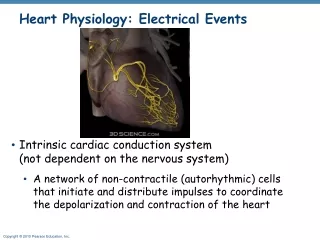
Heart Physiology: Electrical Events Intrinsic cardiac conduction system A network of noncontractile (autorhythmic) cells that initiate and distribute impulses to coordinate the depolarization and contraction of the heart
Autorhythmic Cells Have unstable resting potentials (pacemaker potentials or prepotentials) due to open slow Na+ channels At threshold, Ca2+ channels open Explosive Ca2+ influx produces the rising phase of the action potential Repolarization results from inactivation of Ca2+ channels and opening of voltage-gated K+ channels
Threshold Action potential 2 2 3 1 1 Pacemaker potential 3 2 1 Repolarization is due to Ca2+ channels inactivating and K+ channels opening. This allows K+ efflux, which brings the membrane potential back to its most negative voltage. Depolarization The action potential begins when the pacemaker potential reaches threshold. Depolarization is due to Ca2+ influx through Ca2+ channels. Pacemaker potential This slow depolarization is due to both opening of Na+ channels and closing of K+ channels. Notice that the membrane potential is never a flat line. Figure 18.13
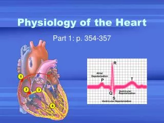
Superior vena cava Right atrium Thesinoatrial (SA) node(pacemaker) generates impulses. 1 Internodal pathway Left atrium 2 The impulses pause (0.1 s) at the atrioventricular (AV) node. Purkinje fibers Theatrioventricular (AV) bundle connects the atria to the ventricles. 3 Thebundle branches conduct the impulses through the interventricular septum. 4 Inter- ventricular septum ThePurkinje fibers depolarize the contractile cells of both ventricles. 5 (a) Anatomy of the intrinsic conduction system showing the sequence of electrical excitation Figure 18.14a
Heart Physiology: Sequence of Excitation Sinoatrial (SA) node (pacemaker) Generates impulses about 75 times/minute (sinus rhythm) Depolarizes faster than any other part of the myocardium
Heart Physiology: Sequence of Excitation Atrioventricular (AV) node Smaller diameter fibers; fewer gap junctions Delays impulses approximately 0.1 second Depolarizes 50 times per minute in absence of SA node input
Heart Physiology: Sequence of Excitation Atrioventricular (AV) bundle (bundle of His) Only electrical connection between the atria and ventricles
Heart Physiology: Sequence of Excitation Right and left bundle branches Two pathways in the interventricular septum that carry the impulses toward the apex of the heart
Heart Physiology: Sequence of Excitation Purkinje fibers Complete the pathway into the apex and ventricular walls AV bundle and Purkinje fibers depolarize only 30 times per minute in absence of AV node input
Homeostatic Imbalances Defects in the intrinsic conduction system may result in Arrhythmias: irregular heart rhythms Uncoordinated atrial and ventricular contractions Fibrillation: rapid, irregular contractions; useless for pumping blood
Homeostatic Imbalances Defective SA node may result in Ectopic focus: abnormal pacemaker takes over If AV node takes over, there will be a junctional rhythm (40–60 bpm) Defective AV node may result in Partial or total heart block Few or no impulses from SA node reach the ventricles Treated with installation of Artificial Pacemaker
Extrinsic Innervation of the Heart Heartbeat is modified by the ANS Cardiac centers are located in the medulla oblongata Cardioacceleratory center innervates SA and AV nodes, heart muscle, and coronary arteries through sympathetic neurons Cardioinhibitory center inhibits SA and AV nodes through parasympathetic fibers in the vagus nerves
Electrocardiography Electrocardiogram (ECG or EKG): a composite of all the action potentials generated by nodal and contractile cells at a given time Three waves P wave: depolarization of SA node QRS complex: ventricular depolarization T wave: ventricular repolarization
QRS complex Sinoatrial node Ventricular depolarization Ventricular repolarization Atrial depolarization Atrioventricular node S-T Segment P-Q Interval Q-T Interval Figure 18.16
R Depolarization SA node Repolarization T P Q S 1 Atrial depolarization, initiated bythe SA node, causes the P wave. Figure 18.17, step 1
R Depolarization SA node Repolarization T P Q S 1 Atrial depolarization, initiated bythe SA node, causes the P wave. R AV node T P Q S 2 With atrial depolarization complete,the impulse is delayed at the AV node. Figure 18.17, step 2
R Depolarization SA node Repolarization T P Q S 1 Atrial depolarization, initiated bythe SA node, causes the P wave. R AV node T P Q S 2 With atrial depolarization complete,the impulse is delayed at the AV node. R T P Q S 3 Ventricular depolarization beginsat apex, causing the QRS complex.Atrial repolarization occurs. Figure 18.17, step 3
Depolarization Repolarization R T P Q S 4 Ventricular depolarization iscomplete. Figure 18.17, step 4
Depolarization Repolarization R T P Q S 4 Ventricular depolarization iscomplete. R T P Q S 5 Ventricular repolarization beginsat apex, causing the T wave. Figure 18.17, step 5
Depolarization Repolarization R T P Q S 4 Ventricular depolarization iscomplete. R T P Q S 5 Ventricular repolarization beginsat apex, causing the T wave. R T P Q S 6 Ventricular repolarization iscomplete. Figure 18.17, step 6
Depolarization Repolarization SA node R R T P T P Q S 1 Atrial depolarization, initiatedby the SA node, causes theP wave. Q S 4 Ventricular depolarizationis complete. R AV node R T P T P Q S Q 2 With atrial depolarizationcomplete, the impulse isdelayed at the AV node. S 5 Ventricular repolarizationbegins at apex, causing theT wave. R R T P T P Q S Q S 3 Ventricular depolarizationbegins at apex, causing theQRS complex. Atrialrepolarization occurs. 6 Ventricular repolarizationis complete. Figure 18.17
(a) Normal sinus rhythm. (b) Junctional rhythm. The SA node is nonfunctional, P waves are absent, and heart is paced by the AV node at 40 - 60 beats/min. (d) Ventricular fibrillation. These chaotic, grossly irregular ECG deflections are seen in acute heart attack and electrical shock. (c) Second-degree heart block. Some P waves are not conducted through the AV node; hence more P than QRS waves are seen. In this tracing, the ratio of P waves to QRS waves is mostly 2:1. Figure 18.18
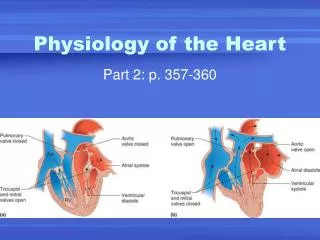
Mechanical Events: The Cardiac Cycle Cardiac cycle: all events associated with blood flow through the heart during one complete heartbeat Systole—contraction Diastole—relaxation
Phases of the Cardiac Cycle • Ventricular filling—takes place in mid-to-late diastole • AV valves are open • 80% of blood passively flows into ventricles • Atrial systole occurs, delivering the remaining 20% • End diastolic volume (EDV): volume of blood in each ventricle at the end of ventricular diastole
Phases of the Cardiac Cycle • Ventricular systole • Atria relax and ventricles begin to contract • Rising ventricular pressure results in closing of AV valves • Isovolumetric contraction phase (all valves are closed) • In ejection phase, ventricular pressure exceeds pressure in the large arteries, forcing the SL valves open • End systolic volume (ESV): volume of blood remaining in each ventricle
Phases of the Cardiac Cycle • Isovolumetric relaxation occurs in early diastole • Ventricles relax • Backflow of blood in aorta and pulmonary trunk closes SL valves and causes dicrotic notch (brief rise in aortic pressure)
Left heart QRS P T P Electrocardiogram 1st 2nd Heart sounds Dicrotic notch Pressure (mm Hg) Aorta Left ventricle Atrial systole Left atrium EDV Ventricular volume (ml) SV ESV Atrioventricular valves Open Closed Open Aortic and pulmonary valves Closed Open Closed Phase 1 2a 2b 3 1 Left atrium Right atrium Left ventricle Right ventricle Ventricular filling Atrial contraction Ventricular ejection phase Isovolumetric relaxation Ventricular filling Isovolumetric contraction phase 1 2a 2b 3 Ventricular filling (mid-to-late diastole) Ventricular systole (atria in diastole) Early diastole Figure 18.20
Cardiac Output (CO) Volume of blood pumped by each ventricle in one minute CO = heart rate (HR) x stroke volume (SV) HR = number of beats per minute SV = volume of blood pumped out by a ventricle with each beat
Cardiac Output (CO) At rest CO (ml/min) = HR (75 beats/min) SV (70 ml/beat) = 5.25 L/min Maximal CO is 4–5 times resting CO in nonathletic people Maximal CO may reach 35 L/min in trained athletes Cardiac reserve: difference between resting and maximal CO
Regulation of Stroke Volume SV = EDV – ESV Three main factors affect SV (stroke volume) Preload Contractility Afterload
Regulation of Stroke Volume Preload: degree of stretch of cardiac muscle cells before they contract (Frank-Starling law of the heart) Cardiac muscle exhibits a length-tension relationship At rest, cardiac muscle cells are shorter than optimal length Slow heartbeat and exercise increase venous return Increased venous return distends (stretches) the ventricles and increases contraction force
Regulation of Stroke Volume Contractility: contractile strength at a given muscle length, independent of muscle stretch and EDV Positive inotropic agents increase contractility Increased Ca2+ influx due to sympathetic stimulation Hormones (thyroxine, glucagon, and epinephrine) Negative inotropic agents decrease contractility Acidosis Increased extracellular K+ Calcium channel blockers
Regulation of Stroke Volume Afterload: pressure that must be overcome for ventricles to eject blood Hypertension increases afterload, resulting in increased ESV and reduced SV In order to compensate, the heart must work harder to overcome the increased blood pressure
1. Which of the following hormones is manufactured by the hypothalamus? (MACA, pick 3) A) Growth Hormone Releasing Hormone B) Growth Hormone Inhibiting Hormone C) Thyrotropine Releasing Hormone D) Thyroid Stimulating Hormone E) hGH
2. Which of the following hormones are released by the ANTERIOR Pituitary Gland? (MACA, pick 2) A) Oxytocin B) Prolactin C) Thyroid Stimulating Hormone D) Anti Diuretic Hormone E) Thyrotropin Inhibiting Hormone
3. Which of the following special senses travels via the Oculomotor Nerve? A) Vision B) Hearing C) Smell D) Taste E) None of the Above
4. Formation of Red Blood Cells is known as... A) You jerk, we just talked about this today! B) We haven't studied this yet C) When in doubt, go with C? Maybe? D) Erythropoiesis E) Not the Answer
5. Which of the following papillae found on the tongue contain taste buds? (MACA, Pick 3) A) Filiform B) Fungiform C) Lentiform D) Circumvallate E) Foliate
6. The functional receptors responsible for dynamic equilibrium is/are the _________ located in the semicircular canals. A) Saccule B) Utricle C) Ampullae D) Crista Ampullaris E) All of the above
7. The “blind spot” of the retina is found where? A) The Fundus B) The Macula Lutea C) The Fovea Centralis D) Two of the above E) None of the above
8. A 45y/o male patient presents for a routine eye examination. A fundoscopic examination reveals that their retina is mildy distorted; the arteries and veins appear to be slightly tortuous. The patient's peripheral vision is decreased in all planes, but the patient has no change in sharp visual acuity. Blood pressure is 122/78 and blood/glucose is 108mg/dL. Urinalysis is – for glucosuria or proteinuria. Which of the following is the most likely diagnosis for the patient's loss of peripheral vision, and which structure is involved? A) Diagnosis: Prehypertension. Structure: Optic chiasm B) Diagnosis: Diabetes Mellitus. Structure: Retina C) Diagnosis: Glaucoma. Structure: Vitreous humor D) Diagnosis: Macular Degeneration. Structure: Macula Lutea E) Diagnosis: Glaucoma. Structure: Canal of Schlemm
9. That last question was hard. Which of the following fat soluble vitamins is the precursor to Retinal? A) Vitamin K B) Vitamin A C) Vitamin D D) Vitamin E E) None of the above