Heart Physiology
Heart Physiology. Physiology of Heart. Heart muscle cells contract, without nerve impulses, in a regular, continuous way Heart is autorhythmic Initiate, conduct and impulse Heart contains special tissue that produces & sends electrical impulses to the heart muscle to contract.

Heart Physiology
E N D
Presentation Transcript
Physiology of Heart Heart muscle cells contract, without nerve impulses, in a regular, continuous way Heart is autorhythmic Initiate, conduct and impulse Heart contains special tissue that produces & sends electrical impulses to the heart muscle to contract
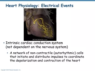
Autorhythmic cardiac cells are found in the following areas: Sinoatrial (SA) node Atrioventricular (AV)node Bundle of His Bundle branches Purkinje fibers Physiology of Heart
Sinoatrial (SA) Node Electrical impulse that causes rhythmic contraction of heart muscles arises in the SA node Located in R. atrium Pacemaker of the heart Generates impulses 70 to 80 times a minute The electrical impulse from the SA node spreads over the right and left atria Causes atrial contraction Physiology of Heart
Physiology of Heart AV Node: Then impulses are conducted to the atrioventicular (AV) node Impulse is delayed at AV node for 0.1 sec Allows completion of atrial contraction before venticular contraction begins
Bundle of His (AV bundle) Then electrical impulse is relayed down to Bundle of His Bundle of His passes impulse to right and left bundle branches Bundle Branches Right and left Bundle branches Branch into purkinje fibers Physiology of Heart
Cardiac Conduction System • Purkinje Fibers • Enter myocardium of ventricle walls, and apex of the heart • Purkinje fibers transmit the impulse first to apex of the heart • Contraction begins at apex and pushes the blood to aorta and pulmonary trunk
CARDIAC CONDUCTION SYSTEM SUMMARY Sinoatrial Node AV Node AV Bundle Bundle Branches Purkinje Fibers
Action Potential in Heart Cells • Conducted cell to cell • Takes 200-500 ms to complete • Resting membrane potential is • electronegative • Unstable resting membrane potential • Continuously depolarize • AP takes place in SA node • When spontaneously changing • potentials, called prepotential • reaches threshold
Action Potential in Heart Cells • Voltage gated Na+ channels • open, Na + influx, K+ channels • close • Depolarization takes place • Depolarized to +20 mV • Repolarization takes place • Na+ channels close, K+ channels • open
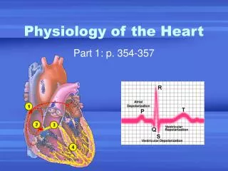
Electrocardiogram: ECG • Conduction of action potential produces • electric current that can be measured • at the surface of the body • P wave: Atrial depolarization • QRS complex: Ventricular • depolarization • T wave: Ventricular • repolarization
Alterations in an Electrocardiogram Normal SA Node Dysfunction no P waves Ventricular Fibrillation
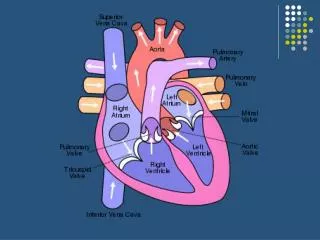
Cardiac Cycle • Heart is two pumps that work together, right and left half • Each pump consists of • Primer pump – Atrium • Power pump – Ventricle
Cardiac Cycle Cardiac cycle: Is the sequence of events in one heartbeat It is the repetitive pumping process that begins with onset of cardiac muscle contraction and ends with beginning of next contraction Cardiac muscle contraction is responsible for pressure and blood movement. How? Blood moves from high pressure to low pressure
Cardiac Cycle The length of cardiac cycle is about 0.8 sec Interval from end of one contraction to the following contraction Consists of Two Phases: Systole phase Diastole phase
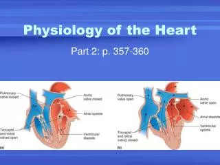
CARDIAC CYCLE • Systole Phase • Contraction phase • Blood ejected • Atrial Systole (0.1 sec.) • Following passive filling with blood • Atrial pressure rises above ventricular pressure • And AV valves open, semilunar valves closed • Ventricles fill with blood semilunar valves (closed) LA bicuspid (open) RA LV tricuspid (open) RV
CARDIAC CYCLE semilunar valves (open) • Systole Phase (cont.) • Ventricular Systole (0.3 sec.) • AV and semilunar valves closed until pressure opens semilunar valves • Blood pushed into pulmonary trunk and Aorta • 120 mm Hg pressure • Atria in diastole LA bicuspid (closed) RA LV tricuspid (closed) RV
CARDIAC CYCLE semilunar valves (closed) • Diastole Phase • Relaxation phase • Ventricular Diastole • Follows ventricular systole • AV valves reopen and filling begins • 80 mm Hg pressure LA bicuspid (open) RA LV tricuspid (open) RV
Heart Sounds • First heart sound or “lubb” • Atrioventricular valves and surrounding fluid vibrations as valves close at beginning of ventricular systole • Second heart sound or “dupp” • Results from closure of aortic and pulmonary semilunar valves at beginning of ventricular diastole, lasts longer
Mean Arterial Blood Pressure • BP is important for blood movement • Blood flows from higher to lower pressure • During one cardiac cycle, blood flows from high pressure in aorta from contraction of ventricles • Then towards the lower pressure in relaxed R. atrium • Mean Arterial Pressure (MAP) = CO x PR • CO (Cardiac output) is amount of blood pumped by heart per minute • PR (Peripheral resistance) is total resistance against which blood must be pumped
CO = HR x SV HR: Heart rate (number of times heart beats per minute) SV: Stroke volume (blood pumped during each heart beat) CO = 72 bpm X 70 ml/beat = 5040ml/min (app. 5L/min) Starling’s law of the heart— the more the cardiac muscle is stretched, the stronger the contraction Important factor for stretching the heart muscle is venous return Greater the volume of blood returned to the heart by the veins, Greater the volume of blood the heart will pump Cardiac Output
Regulation of the Heart • To maintain homeostasis, amount of blood pumped by heart must vary: • Eg. Cardiac output increases more during exercise than resting • Intrinsic regulation: Results from normal functional characteristics of heart, not depend on neural or hormonal regulation
Regulation of the Heart • Extrinsic regulation: Involves neural and hormonal control • Neural Control • Parasympathetic stimulation • Supplied by vagus nerve, acetylcholine is secreted, • decreases heart rate, maintain • heart beat average of 70 beats/min. • Sympathetic stimulation • Supplied by cardiac nerves • Increases heart rate and force of contraction. • Epinephrine and norepinephrine released. • Increased heart beat causes increased cardiac output
Regulation of the Heart Hormonal Control Epinephrine and norepinephrine from the adrenal medulla Increases rate and force of heart contraction Occurs in response to increased physical activity, emotional excitement, stress
Heart and Homeostasis • Effect of blood pressure • Baroreceptors monitor blood pressure; in walls of internal carotids and aorta. This sensory information goes to centers in the medulla oblongata
Heart and Homeostasis • Effect of pH, carbon dioxide, oxygen • Receptors that measure pH and carbon dioxide levels found in hypothalamus • Chemoreceptors monitoring oxygen levels found in aorta and internal carotids. Prolonged lowered oxygen levels causes increased heart rate, which increases blood pressure and can thus deliver more oxygen to the tissues.
Heart and Homeostasis • Effect of extracellular ion concentration • Increase or decrease in extracellular K+ decrease the heart rate • Effect of body temperature • Heart rate increases when body temperature increases, heart rate decreases when body temperature decreases
Effects of Aging on the Heart • Gradual changes in heart function, minor under resting condition, more significant during exercise • Hypertrophy of left ventricle • Maximum heart rate decreases • Increased tendency for valves to function abnormally • Increased oxygen consumption required to pump same amount of blood
DISORDERS Tachycardia Abnormally high heart rate (over 100) Bradycardia Abnormally low heart rate (under 60) Fibrillation Rapid and out of phase contractions Atherosclerosis Formation of fatty plaque on artery walls Decrease in vessel elasticity and possible blockage
Dynamics of Blood Circulation
Dynamics of Blood Circulation • Interrelationships between • Pressure • Flow • Resistance • And the control mechanisms that regulate blood pressure and blood flow • Play important role in circulatory system
Laminar and Turbulent Flow • Laminar flow • Blood flow in Streamlined fashion • Interior of blood vessel is smooth and of equal diameter along its length • Outermost layer moving slowest (move against resistance of stationary wall) • And center layer moving fastest
Laminar and Turbulent Flow • Turbulent flow • Interrupted • Fluid passes a constriction, sharp turn, rough surface • Partially responsible for heart sounds • Sounds due to turbulence not normal in arteries and is probably due to some abnormal constriction • And increases the probability of thrombosis
Blood Pressure • Blood pressure: Measure of force exerted by blood against the blood vessel wall • Blood moves through vessels because of blood pressure • BP is measured in mm Hg • Measured by Sphygmomanometer • Measured by listening for Korotkoff sounds produced by turbulent flow in arteries as pressure released from blood pressure cuff (systolic pressure) • No sound, continuous laminar flow, Diastolic pressure
Pulse Pressure • Pulse Pressure: Difference between systolic and diastolic pressures • Healthy person 120 mm Hg systolic, 80 mm Hg diastolic • Pulse pressure is 40 mm Hg • Pulse pressure increases when stroke volume increases • eg. During exercise, stroke volume increases, pulse pressure also increases • Pulse pressure can be used to take a pulse to determine heart rate • Most frequent site used to measure pulse rate is in the carpus with the radial artery- the radial pulse
Blood Flow • Rate of flow through a tube is expressed as the volume that passes a specific point per unit of time. E.g.; cardiac output at rest is 5L/min, thus blood flow through the aorta is 5L/min • Blood flow = (P1 – P2/R) • P1 and P2 are pressures in the vessel at points one and two; R is the resistance to flow • Blood flow is directly proportional to pressure differences, inversely proportional to resistance • Resistance = 8vl/r4 • v is viscosity, l = length of the vessel, r is the radius of the vessel, 8 and are constant • radius and viscosity determines resistance
Poiseuille’s Law • Blood flow decreases when resistance increases • Since resistance is proportional to blood vessel diameter, constriction of a blood vessel increases resistance and thus decreases flow • Blood Flow =(P1 –P2)/R • Poiseuille`s law = (P1 –P2)r4/8vl • According to Poiseuille`s law, small change in radius dramatically changes resistance to flow, r is raised to power 4 • During exercise, blood vessels in skeletal muscle vasodilate, decreases resistance to blood flow • And blood flow through blood vessels increases dramatically
Viscosity • Viscosity: Is measure of resistance of liquid to flow • As viscosity increases, pressure required to flow increases • Viscosity of blood is influenced largely by hematocrit (percentage of the total blood volume composed of red blood cells) • Dehydration and/or uncontrolled production of RBCs can increase hematocrit, thus increase viscosity • Higher viscosity increases the workload on the heart, heart failure can result
Influences on B.P. • Blood Pressure varies directly with the following: • Blood Volume • Mainly regulated by kidneys • in blood volume = in B.P. • in blood vol. = decrease in B.P.
Regulation of Blood Pressure • By nervous system, kidneys and chemical controls • Nervous System Regulation: • Sympathetic nerve fibers: • Vasoconstriction of blood vessels • diameter, resistance B.P. • Vasomotor center in medulla: • Controls cardiac output • Controls degree of vessel constriction
Chemical Regulation of Blood Pressure • Epinephrine and Norepinephrine • - Vasoconstriction • - cardiac output, B.P. • ANF (Atrial Natriuretic Factor) • - ANF act on kidney • - Release of more sodium and • water in urine • - Loss of water and sodium in urine • - blood volume B.P.
Chemical Regulation of Blood Pressure • ADH (Antidiuretic Hormone) • - Stimulates kidneys to reabsorb water • - blood volume B.P. • Renin (Enzyme) • - Released from kidneys in response to low • B.P. • - Stimulates angiotensin/aldosterone system • - Kidneys reabsorb sodium and water • blood volume and B.P.
RENAL REGULATION OF B.P. • Kidneys may alter B.P. directly • - Increased B.P. more blood filtered by kidneys • - More urine produced and released • - blood volume B.P. • Kidneys may alter B.P. indirectly • - Renin/angiotensin system activated with B.P. • - Vasoconstriction, water reabsorption due to • aldosterone release • - blood volume B.P