Restrictive Cardiomyopathy
Restrictive Cardiomyopathy. Loryn S. Feinberg Wednesday, April 20, 2005 Echocardiography Conference. Restrictive Cardiomyopathy. Definition Characteristics Clinical Presentation Classification Specific Diseases & Echo Findings General Echo Features. Definition.

Restrictive Cardiomyopathy
E N D
Presentation Transcript
RestrictiveCardiomyopathy Loryn S. Feinberg Wednesday, April 20, 2005 Echocardiography Conference
Restrictive Cardiomyopathy • Definition • Characteristics • Clinical Presentation • Classification • Specific Diseases & Echo Findings • General Echo Features
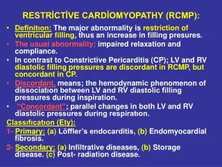
Definition • Idiopathic or systemic myocardial disease characterized by: • Impaired ventricular filling • Elevated diastolic pressures • Normal or reduced diastolic volume of ventricle(s) • Normal/near normal systolic function until advanced stages
Clinical & Echo Characteristics • Nondilated ventricle • Normal to increased wall thickness • Rigid ventricular walls • Severe diastolic dysfunction • Restrictive filling • Elevated diastolic filling pressures • Dilated atria, elevated RA pressure • Pulmonary hypertension • Inability to ↑ CO with exercise due to impaired filling • Right sided failure Benotti, JR et al. Clinical profile of restrictive cm. Circ 80’ 61:1206
Clinical Presentation • Signs of pulmonary and systemic congestion in absence of cardiomegaly • Dyspnea • PND, orthopnea • Peripheral edema • Palpitations • Fatigue, weakness, exercise intolerance • Thromboembolic complications (up to 1/3 with idiopathic RCM) • Cardiac conduction disturbances • Amyloid , sarcoid, hemochromatosis • AF common in IRCM & amyloidosis • Advanced Stage • Marked elevation in CVP • Hepatosplenomegaly, ascites, anasarca
Physical Examination • Pulse: normal or low amplitude/tachycardic [low SV] • JVP: elevated with prominent x and y descents • Kussmaul’s sign: JVP fails to fall or ↑es w/ inspiration • Increased resistance to RA filling during inspiration • LV impulse: normal • S1, S2 normal, often S3 present • abrupt cessation of rapid ventricular filling • Regurgitant murmurs • Peripheral edema, ascites, pulsatile liver, HSM
Differentiation from Constrictive Pericarditis Adapted from Hurst’s The Heart, 10th ed. 2004
Secondary restrictive physiology may occur in advanced stages of dilated, hypertrophic, hypertensive, ischemic heart disease Kushwaha et al. 336 (4): 267, Table 1 January 23, 1997
Myocardial, Non-infiltrative • Idiopathic • Sporadic , AD, or AR • Familial type: part of spectrum of familial HCM? • Different phenotypic expression of same genetic disease • May be associated with distal skeletal myopathy, occasionally heart block [fibrosis of SA & AV nodes] • Manifests at any age [childrens’ prognosis worse, adults’ course protracted] • Incidence in elderly, women men • Survival time variable, mean 9 years • Disease of exclusion Marked patchy interstitial fibrosis
Idiopathic Restrictive CM:2D Echo features • Biatrial enlargement • Thrombi in atrial appendages • Cavity size/wall thicknessnormal • Normal or reduced global systolic function • Right ventricle eventually enlarges [depending on degree of PH] • May have patchy, granular sparkling appearance Non-dilated, non-hypertrophied ventricles with dilated atria
Myocardial, Non-infiltrative • Scleroderma • Myocardial fibrosis, contraction band necrosis [dense bands through myofibers often seen after ischemiai w. reperfusion] • Often patchy, may be biventricular • Microvascular occlusions → ischemia • Fibrinous pericarditis, effusions • Ventricular conduction abnormalities • Heart block, SVTs, VT, pseudoinfarction patterns on ECG • Pulmonary hypertension leading cause of morbidity/mortality Fibrous tissue replacement: Thinned papillary muscles & LV wall Braunwald, Heart Disease; 6th edition
Myocardial, Infiltrative • Amyloid • Primary:light chain immunoglobulin overproduction from monoclonal plasma cells (multiple myeloma) • 50% clinical cardiac involvement • Secondary:fragments of serum amyloid A protein • Chronic inflammatory conditions (Crohn’s, RA, Tb, FMF) • 10% clinical cardiac involvement • Familial & Senile: overproduction of transthyretin [>50 mutations] • Familial usually AD, associated with ascending peripheral & autonomic neuropathy • ‹ 5% clinical cardiac involvement Dx: endomyocardial or fat biopsy Left: prominent interstitium, expansion by acellular, eosinophilic substance. [Uneven size myocardial cells, vacuolated] Right: affinity for sulfated alcian blue (histochemical equivalent of congo red stain)
Myocardial, Infiltrative • Amyloid • Deposits may be interstitial & widespread: • Myocardial Dysfunction: • Diffuse infiltration of myocardium with stiff beta-pleated fibrils →impaired relaxation, diastolic dysfunction • Replacement of functional myocardium with amyloid-> systolic dysfunction • Stiff Heart Syndrome: restrictive filling present & impaired systolic function • Deposits may localize to: • Conduction tissue: heart block, ventricular arrhythmias • Valves: regurgitation • Pericardium: constriction • Coronaries: ischemia • Prognosis poor, median survival two years • Diastolic function found to be a stronger predictor of cardiac death than LV wall thickness or systolic function • Cardiac transplantation not usually performed Swanton, RH et al. Systolic and diastolic ventricular function in cardiac amyloidosis. Am J Cardiol 1977; 39:658. Utility of echocardiography in evaluation of individuals with cardiomyopathy. Heart 2004;90
Amyloid: 2D Echo features • Ventricular cavities may be small, normal, or moderately dilated • Atrial appendage thrombi • Dilated atria & IVC • Normal or increased wall thickness • Prognostic variable • Survival range 2.4 yrs if NL(≤12 mm) • 0.4 yrs if markedly (15 mm) • Variable (but often depressed) systolic function • Involvement of pericardium, valves, coronaries • Granular, sparkling appearance is characteristic BiV hypertrophy, biatrial enlargement, & mild, diffuse valve thickening Cueto-Garcia L, et al. Echo findings in systemic amyloidosis. JACC 85; 6 Katritsis, D et al. Primary restrictive CM: clinical and pathologic characteristics. JACC 91; 18
Myocardial, Infiltrative • Sarcoidosis • Affects young/middle age adults, no gender prediliction • Noncaseating granulomas in lungs, spleen, lymph nodes, skin, liver, parotid glands, heart • Infiltration of conduction system, LV (upper septum), pulmonary artery • Autopsy: Cardiac involvement in 25%, Clinically: 5% • Cardiac manifestations: restrictive cm->dilated • Conduction abnormalities, high-grade AV block, VT • Patchy distribution • Biopsy sensitivity of 20-30% • Sudden cardiac death in 17% with extensive cardiac granulomas • Cardiac transplantation may be appropriate for intractable heart failure or arrhythmias Griffin BP; Manual of CV Medicine 2004
Sarcoidosis: Echo features • Systolic function usually normal initially • Diffuse HK & focal abnormalities of WM • Basal septum affected, apex spared • Pulmonary involvement frequent • Pulmonary hypertension, RHF • LV aneurysms • Valvular regurgitation • Septum or LV free wall may appear hyperechogenic Valantine H et al. Sarcoidosis: a pattern of clinical and morphological presentation. Br Heart J 87’ 57 Fahy, J et al. Doppler echo detection in patients with pulmonary sarcoidosis. Chest 96’ 109
Myocardial, Infiltrative • Gaucher’s • Most common lysosomal storage disease • Deficiency in beta-glucocerebrocidase enzyme • Accumulation of cerebroside in many organs, usually bm, spleen, liver, brain • More rare: pulmonary & cardiovascular involvement • Cerebroside accumulation in interstitium of LV -> restrictive cm • Often subclinical • Pulmonary hypertension from pulmonary capillary occlusion • Calcification & thickening of valves, pericardial effusion • Hurler’s Syndrome • Mucopolysaccharide accumulation leads to severe skeletal deformities, hepatosplenomegaly, mental retardation • Cardiac involvement evident between 1-5 years of age • Mucopolysaccharide deposition in myocardial interstitium, valves, coronary arteries, aorta
Metabolic StorageDiseases • Hemochromatosis • Five types, AD or AR • Iron deposition in multiple organs (heart, liver, skin, pancreas, pituitary, joints) • Dilated>restrictive cardiomyopathy • Systolic dysfunction indicates a poor prognosis • Conduction disturbances common, e.g., SSS • SVT & VT occur in 1/3 of patients • Granular or sparkling appearance may be seen on echo • Atrial enlargement • Treatment with repeated phlebotomy or chelation (desferrioxamine) may reverse LV dysfunction • Cardiac transplantation can be considered Hurst’s The Heart, 10th edition
Fabry Disease (angiokeratoma corporis diffusum,ceramide trihexosidosis, or Anderson-Fabry disease ) • X-linked disorder of glycosphingolipid metabolism • Due to deficiency of lysosomal ceramide (- galactosidase) • Intracellular accumulation & deposition of glycolipids in heart, skin, kidneys • First symptoms manifest by age 10 • Peripheral neuropathy, hypohidrosis, skin lesions (angiokeratomas), renal dysfunction Nagueh, SF. Fabry disease. Heart 2003; 89:819.
Fabry Disease • Accumulation occurs in myocardium, vascular, and valvular endothelium • May develop LVH (concentric) with hypertrophic CM, restrictive CM, or dilated CM • Conduction abnormalities, AR>MR, premature CAD, aortic root dilation, premature stroke • Cardiac manifestations may not occur until age 30 • Echo findings are similar to amyloid • LV mass correlates with disease severity • Full expression in males, incomplete in females • Screening for plasma alpha-galactosidase A levels in patients with unexplained concentric LVH should be considered • Recombinant alpha-galactosidase causes regression of cardiac dysfunction
Endomyocardial Diseases Endomyocardial Fibrosis (Davies Disease) • Tropics • 10-20% of deaths due to HF in equitorial Africa • Children, young adults • No gender prediliction • Intense endocardial fibrotic thickening of apex & subvalvular regions of ventricle(s)→ obstruction to blood inflow, regurgitation, restrictive physiology • Histology: granulation tissue, collagen, connective tissue lines endocardium • Affects both (50%), left (40%), right (10%) ventricles • Two year mortality up to 50%
Endomyocardial Fibrosis:Echo Features • Inferobasal LV wall thickening • Endocardial thrombus deposition • Apical obliteration • Biventricular involvement in ½ of patients • Ventricular cavities vary in size but often obliterated by extensive endocardial thickening • Large atria • Papillary muscle involvement & AV valve apparatus with valve deformity common • Regurgitation>stenosis • AF common
Endomyocardial Diseases • Loeffler’s endocarditis [eosinophilic cardiomyopathy] • Temperate climates • Activated eosinophils may release cardiotoxic intracytoplasmic granules • Middle age, usually men • Intense eosinophilia, nervous system & bone marrow involvement, skin rash, constitutional symtpms • Restrictive cardiac disease (often biventricular), fibrinoid vasculitis of coronaries • Valvular involvement leads to regurgitation or stenosis of AV valves • Thromboembolic phenomena • Mural & atrial thrombi • LV thrombi in absence of WMA • Gradual apical obliteration; apex fills with echogenic mass Tai PC Lancet 87’; 1; Wood MJ et al; Utility of Echo in the Eval of Ind with CM; Heart 2004; 90 Thrombus deposition in RV/LV apices
Endomyocardial Diseases • Carcinoid Heart Disease • Late complication of carcinoid syndrome from metastatic carcinoid tumors • 50% of patients • TR most common finding • Pathognomonic fibrous plaque-like deposits • Pulmonary inactivation of serotonin, bradykinin, etc.. • LV involvement uncommon • If present, likely r->l shunt • Endocardium of valve cusps/leaflets,cardiac chambers, intima of PA/aorta • Plaques: smooth muscle cells embedded in mucopolysaccharides & collagen • Lack elastic components Pellikka PA et al. Carcinoid heart disease; clinical and echo spectrum in 74 patinets.Circ 93’ 87
Carcinoid Syndrome • Echo findings in 2/3 of patients • Thickened, retracted, immobile tricuspid & pulmonary valves • RA/RV enlargement • RA wall thickening on TEE • Pulmonary outflow obstruction may occur • TR/PR on doppler • Rarely see metastatic tumors to heart • Valvular lesions do not regress with therapy
Endomyocardial Diseases • Radiation carditis • Can manifest decades after thoracic radiotherapy • Can involve many cardiac structures • Coronary arteries: ostial stenoses • Valves: stenosis • Pericardium: constrictive pericarditis • Myocardium, endocardium: • Interstitial fibrosis of RV > LV Brosius FC III et al. Radiation heart disease. Am J Med 81’; 70.
EndomyocardialDiseases • Metastatic cancer (lung, breast, melanoma, leukemia, lymphoma) • Drug toxicity: • Anthracylines: dilated CM & endomyocardial fibrosis • Risk of restrictive cm markedly increased with concurrent irradiation • Diastolic abnormalities do not appear to be dose-related • Drugs causing endomyocardial fibrosis • Methylsergide • Serotonin • Busulfan • Ergotamine • Mercurial agents
Doppler Echo findings: Early in Disease Course • Impaired diastolic filling • Doppler of LV inflow • Reduced E velocity, increased A velocity • Prolonged isovolumic relaxation time • Decreased early diastolic deceleration slope • Pulmonary vein inflow • Reduced diastolic filling phase • Normal systolic filling phase • Decreased ratio of systolic to diastolic PV flow Zile et al. Circ 02’; 105
Pseudonormalization • LA pressure rises • Increased pressure gradient from LA to LV at MV opening • E and A velocities may remain relatively normal • Pulmonary venous inflow • Systolic filling normal or decreased • Diastolic is normal or increased • Increased resistance to LV filling : in velocity and duration of atrial flow reversal in lower resistance PV Otto, Textbook of Clinical Echo; 3rd edition
Advanced Restrictive Pattern • Ventricular compliance continues to decrease • Increased mitral early diastolic filling velocity (E) [> 1m/s] • Deceleration slope becomes more rapid • A velocity reduced due to increased LVEDP & reduced atrial contractile function [<.5 m/s] • Increased E:A ratio (>2) • Mitral deceleration time shortens (<150 ms) • Isovolumic relaxation time shortens [ 70 ms] • PV flow shows increased diastolic phase, reduced systolic phase & Pva increases further in duration & velocity E/A 2.4
Myocardial Tissue Doppler Signals • Displays the velocities of the myocardium during contraction and relaxation • Can assist in assessment and grading of diastolic function • Sm: myocardial velocity during systole • Em: myocardial velocity during early filling • Am: myocardial velocity during filling due to atrial contraction • In restrictive pattern, Em is diminutive • Helpful in differentiating constriction from restriction
Cardiac MR • Can aid in distinguishing between the cardiomyopathies • Hemochromatosis produces low signal intensity due to iron deposition • May differentiate primary restrictive CM from amyloidosis based on tissue characterization • May detect mass lesions due to sarcoid granuloma or scar • Can aid in differentiating constrictive from restrictive disease Celetti et al, AJC 99’ 83