Cardiomyopathy
Cardiomyopathy. A disease due to Primary Pathology in the Myocardium i.e. No evidence of significant occlusion of the Coronary Arteries. i.e. No evidence of Ischemic “Cardiomyopathy”. Classification. Dilated (Common) (Systolic Disorder) Hypertrophic (Uncommon) (Diastolic Disorder)

Cardiomyopathy
E N D
Presentation Transcript
Cardiomyopathy A disease due to Primary Pathology in the Myocardium i.e. No evidence of significant occlusion of the Coronary Arteries. i.e. No evidence of Ischemic “Cardiomyopathy”
Classification • Dilated (Common) (Systolic Disorder) • Hypertrophic (Uncommon) (Diastolic Disorder) • Restrictive (Rare) (Both Systolic & Diastolic)
Dilated Cardiomyopathy • Clinical:Any Age Patients present with Arrythmia (Atrial Fib) Congestive Failure Pul. Edema J.V.P. Thromboembolic Disease • Echo Poor contraction of L.V. or R.V. during systole • Causes: Idiopathic Post Viral Alcohol • Familial: Right Ventricular Cardiomyopathy (Italy) Muscular Dystrophy Fredreichs Ataxia
Pathology • Heart heavy – 600 grams + • All 4 chambers dilated & flabby • L.V. wall may appear thin or normal because of dilation of microfibres • L.V. wall 12-15mm thick • Valves – normal • Coronary Arteries – normal • Histology:Hypertrophy – of cardiac nuclei + Interstitial Fibrosis
Hypertrophic Cardiomyopathy H.O.C.M. • Clinical • Sudden Death of a young adult esp. during exercise! • History of “Drop Attacks” esp. during vigorous exercise. • Dyspnoea & Angina – Intramyocardial Coronary Arteries – occluded • Harsh Systolic Murmur
Pathogenesis • Genetic Defect – Hereditary • Autosomal Dominant • 4 genes encode • Proteins of contraction • Myosin genes & Troponin • 50 different mutations • Echo Diagnostic – Septal Hypertrophy + Vigorous Contractions of L.V. Poor Diastolic Filling
Gross • Disproportionate thickening of septum compared to Free Wall – Typical • Microscopic • A – Microfibre Disarray in sections from Septum • B – Massive enlargement of cardiac nuclei
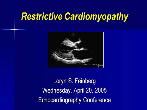
Restrictive Cardiomyopathy • So called because ventricular filling is severely restricted because muscle does not relax during diastole. • Clinical – Because of above – get elevated J.V.P. Pul. Edema and Rt Heart Failure Liver ++ Ankle Edema ++ etc • Echo – Impaired Ventricular filling Normal systole
Causes • Amyloid of Heart • Sarcoid of Heart • Hemochromatosis (Iron storage disease)
Microscopic • Deposits of Amyloid Protein between the microfibres • Diagnosis:- Congo Red Stain – Amyloid Red with Green Briefringence Under polarised light
Amyloid Heart (500 grams) • Gross Laying down of insoluble protein called amyloid between the microfibres • Causes Causes “stiffness” in the tissue and they take on a firm waxy appearance. Heart muscle becomes non-compliant and get failure to Contract & Relax during systole and diastole