Download
1 / 13
140 likes | 494 Vues
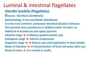
Luminal & intestinal flagellates. G iardia lamblia (flagellate). D isease : Giardiasis ( lambliasis ). Epidemiology: It has worldwide distribution. It is the most common protozoan intestinal disease in disease.

E N D
Luminal & intestinal flagellates Giardialamblia (flagellate). Disease : Giardiasis (lambliasis). Epidemiology: It has worldwide distribution. It is the most common protozoan intestinal disease in disease. This parasite more prevalence in children under 10 years old. Habitat→→duodenum and upper jejunum. Infective stage → → Mature quadrinucleate cyst. Pathogenic stage → →Active trophozoite. Diagnostic stage → → Mature cyst and trophozoite in stool sample. Mode of infection → → Contamination of food and water with cyst. Route of entry → → is mouth or orally.
Morphology: There are two stages: Trophozoite: Morphology: →→ It is 12-15 µ pear shaped with 8 flagella(f) and, 2 axostyles(a) arranged in a bilateral symmetry. →→ There are two anteriorly located large sucking discs(d). →→ The cytoplasm contains two 2 nuclei (n) with karyosome (k) and 2 Para basal bodies(p.b). Cyst: →→ Giardia cysts are 9-12 microns. →→ Ovoid in shape with well-defined cyst wall(c.w). →→ The cytoplasm contains 4 nuclei(n) . →→ many structures of the trophozoite like 2 axostyles (a) and parabasal bodies(p.b).
Life cycle of G. Lamblia →→ Life cycle: Infection occurs by ingestion of cysts, usually in contaminated water. →→ Ex-cystation occurs in duodenum and trophozoites colonize the duodenum where they may swim freely or attach to the sub-mucosal epithelium via the ventral suction disc. →→ The free trophozoites encysted in the colon. →→ The cysts are passed in the stool. →→ Man is the primary host and some animals(e.g. pigs and monkeys, mice…..etc) are also infected and serve as reservoirs. →→ Afterrward, person-to-person transmission is possible.
Symptoms & Pathology: Symptoms: The early symptoms include →→ abdominal pain and distension, nausea and fatty diarrhea (steatorrhea). →→ The stool contains excessive lipids but very rarely any blood or necrotic tissue. The more chronic stage is associated with vitamins A and B12 Malabsorption, disaccharides deficiency. : Pathology →→ Covering of the epithelium by the trophozoite causing malabsorption of nutrients.
Diagnosis Diagnosis: by Symptoms, history, epidemiology. →→ Distinct from other dysentery due to lack of mucus, and blood in the stool, lack of increased PMN leukocytes in the stool and lipids droplets & lack of high fever. →→ Cysts in the stool and trophs in duodenal content obtained using a string device (Enterotest R). →→ Trophozoite must be distinguished from the nonpathogenic flagellate Trichomona hominis, an asymmetrical flagellate with an undulating membrane.
GIARDIA Treatment Treatment: Keep the child hydrated. If it is dehydrated: Using drugs 1-Metronidazole(Flagyle). 2-Tinidazolea new Giardia alternative drugs (but it has a shorter treatment course) used for under 3 years children.
LUMINAL flagellates →→ Lumen-dwelling F: Flagellates found in the lumen of alimentary canal and UGT. Genus :Trichomonas. General characters. →→ The genus Trichomonas consists of 3 main species which infect man. →→ All 3 spps. have direct life cycle . →→ All Trichomonas posses 3 to 5 anterior flagella, an undulating membrane, and a recurrent flagellum attached to the edge of the undulating membrane. →→ An axostyle extended along the length of the body and extends from posterior. →→ reproduce by longitudinal binary division . →→ A cyst stage is not known for this genus.
Trichomonas There are three species of genus Trichomonas 1-Trichomonas tenax →→Living in the mouth and teeth pockets. →→ Moving vigorously by flagellum through saliva, feeding on food debris. →→ The tissue destruction has been done by toxins produced by this and other pathogens at the site of infection. 2-Trichomonas hominis. →→ Lives in the intestine. →→ It is cosmopolitan in distribution. →→ It is thought to be non-pathogenic although it has been associated with diarrheic stools.
3-Trichomonas vaginalis $ $→ → Trichomoniasis disease. $ $→ → It is sexually transmitted disease. Epidemiology: It has a world-wide. Morphology: Trophozoite: half pear shaped with a single nucleus and 4 anterior flagella and a lateral flagellum attached by an undulating membrane, 2 axostyles arranged asymmetrically. The organism does not encysted
Trichomonas vaginalis Pathogenesis: →→ Trophozoite infect the squamous epith. Cells. →→ It is asymptomatic particularly in male. →→ In females causing severe causing irritation& Inflammation of the vaginal mucosa(vaginitis) or inflammation uterine cervix(cervicitis). →→ In males causing inflammation of urethra and prostate (urithritis and prostatitis). Clinical symptoms : →→ majority of infected males without symptoms and ⅓ of infected females without symptoms. →→ both in male and female genital discharge and pain in urination.
Diagnosis and treatment Diagnosis →→ By GUE or males and females UG swab and smear stain with Giemsa stain. Treatment. 1-Metronidazole(Flagyle) tablets for men and vaginal suppositories for females. 2-Tinidazole alternative drug.it has a shorter treatment course.