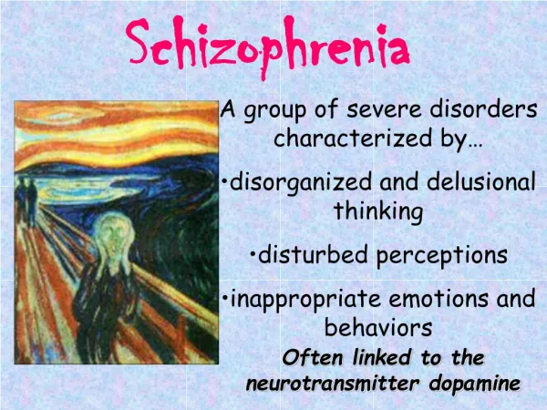
Caudate Shape Discrimination in Schizophrenia
10 likes | 130 Vues
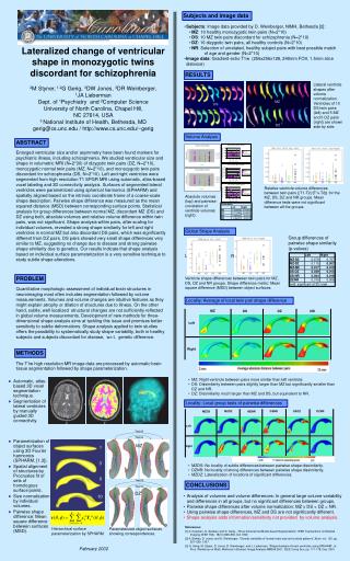
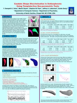
RESULTS. ABSTRACT. SZ vs CNTL. Typ vs CNTL. Atyp vs CNTL. Typ vs Atyp. Left. 0.00083. 0.00001. 0.03037. 0.01493. Right. 0.00398. 0.00002. 0.05228. 0.04111. SHAPE MODELING. CLINICAL STUDY. SZ vs CNTL. Typ vs Atyp. Typ vs CNTL. Atyp vs CNTL. Left. 0.5112. 0.0651. 0.5781.

Caudate Shape Discrimination in Schizophrenia
E N D
Presentation Transcript
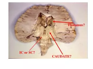
RESULTS ABSTRACT SZ vs CNTL Typ vs CNTL Atyp vs CNTL Typ vs Atyp Left 0.00083 0.00001 0.03037 0.01493 Right 0.00398 0.00002 0.05228 0.04111 SHAPE MODELING CLINICAL STUDY SZ vs CNTL Typ vs Atyp Typ vs CNTL Atyp vs CNTL Left 0.5112 0.0651 0.5781 0.0188** Right 0.5742 0.1518 0.5272 0.07 SZ vs CNTL Typ vs CNTL Typ vs Atyp Atyp vs CNTL Left 0.4326 0.0522 0.7581 0.001** Right 0.4121 0.1202 0.4424 0.0022** STATISTICAL ANALYSIS Caudate Shape Discrimination in Schizophrenia Using Template-free Non-parametric Tests Y. Sampath K. Vetsa1, Martin Styner1, Stephen M. Pizer1, Jeffrey A. Lieberman2, Guido Gerig1,2 1 Department of Computer Science, 2 Department of Psychiatry, University of North Carolina, Chapel Hill, NC 27599, USA We describe the shape analysis of the caudate nucleus structure in a large schizophrenia study. Left and right caudates were segmented from high resolution MRI using a reliable, semi-automated technique. The caudate shapes were parameterized by spherical harmonic surface description, aligned and finally represented as medial mesh structures (m-reps). Schizophrenia patients were categorized based on treatment, so we could test size and shape differences between normals, typically and atypically treated subjects. Statistical shape analysis was done using permutation tests on shapes represented by medial representations. Significant size and shape differences were found, not only between treatment groups and controls, but also between the treatment groups. VOLUME DIFFERENCES For the volume tests, the signed difference between the average volumes of the groups is used as the distance measure, with a a two-sided permutation test. We used 100,000 permutations. Statistics for caudate volumes, with correction for ICV (intracranial volume). The table lists p-values. All comparisons are significant. GLOBAL SHAPE DIFFERENCES The tables below show the m-rep statistics for deformation and width, integrated over the whole shape. The tables list the p-values, with significant results marked with **. We used 10,000 permutation tests with one sided tests. The patient study is cross-sectional in design. Early illness (age 16-30, N=34) and chronic (age 31-60, N=22) patients have been matched to a young and an older control group (N=26). All patients and comparison subjects were right-handed males. At the time of the scan, 17 early illness patients and 5 chronic patients were on typical antipsychotic medications, and 17 early illness and 18 chronic patients were on atypical antipsychotic medications. M-rep statistics for the mesh deformation measure: Mesh deformation distance (L1) calculated as the integrated deformation over the whole mesh. Caudate structures were segmented using a semi-automated technique. Following automatic tissue classification, a trained user masked the gray matter structure of the caudate by overpainting. After segmentation, the shapes were processed by surface extraction and parameterization using spherical harmonics (SPHARM). The shapes were then aligned using first degree spherical harmonics, which also gives the correspondence. An m-rep model is generated with the optimal minimum mesh sampling, and this m-rep model is used to generate the m-reps for the individual objects by deformation into each object. M-rep statistics for the local width measure: Width (radius) change measured as the integrated absolute radius differences over the whole mesh. LOCAL SHAPE DIFFERENCES The two sets of figures below show the local shape difference statistics. In the left figure, the locations on the mesh with p-values more than 0.05 are suppressed, with lower p-value indicated by darker shading. In the right figure, the locations on the mesh with p-values more than 0.05 are suppressed, with the size and color of the ball at each node indicating the significance. Caudate segmentation SPHARM representation PDM + M-rep M-rep + Radius Implied surface Local deformation statistics for Typ-Atyp group tests for the left caudate. The problem with shape analysis is High Dimension Low Sample Size (HDLSS). Possible Approach • Feature Selection • Feature reduction (e.g. Principal Component Analysis and selection of a few modes) • Multivariate statistical analysis Our Approach : Permutation test • Random permutation of shapes • Calculate average shapes • Scalar shape distance measure between shapes • Test significance of the original group average difference Local width statistics for Typ-Atyp group tests for the left caudate. Surfaces implied from m-reps: The figures below show the average m-rep surfaces for the average Atyp (solid) and the average Atyp + Δ(Typ – Atyp)radius overlaid (mesh). Please note that this display differs from an overlay of average shapes and illustrates significant changes (here radius only) between the groups. The figure below shows an example of a permutation test. The number of permutations are on the x-axis, and the scalar shape difference is plotted on the y-axis. The distance between the original groups is plotted as the thick horizontal line. In this case, the difference between the groups is significant. Side View Top View This work was supported by the NCI grant P01 CA47982. The Stanley Medical Research Institute is acknowledged for funding the MRI schizophrenia study providing the caudate structures. Miranda Chakos and Scott Schobel are acknowledged for providing the caudate segmentations.