Download
1 / 119
1.21k likes | 1.5k Vues
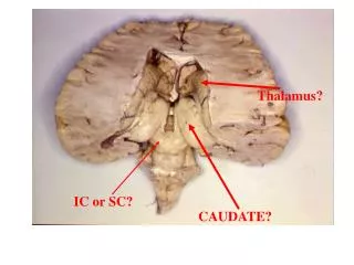
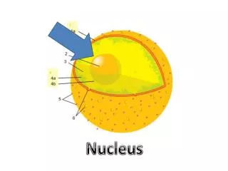
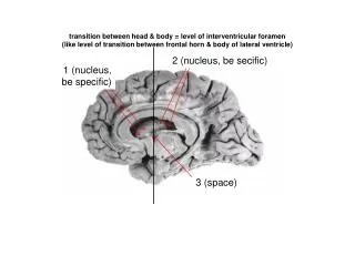
transition between head & body = level of interventricular foramen (like level of transition between frontal horn & body of lateral ventricle). 2 (nucleus, be secific). Body of caudate nucleus. 1 (nucleus, be specific). Head of caudate nucleus. Interventricular foramen. 3 (space).

E N D
transition between head & body = level of interventricular foramen (like level of transition between frontal horn & body of lateral ventricle) 2 (nucleus, be secific) Body of caudate nucleus 1 (nucleus, be specific) Head of caudate nucleus Interventricular foramen 3 (space)
Position of: ventral corticospinal, vestibulospinal, & reticulospinal tracts 2 ventral horn with LMN 5 lateral corticospinal & rubrospinal tracts 4 lateral corticospinal tract + rubrospinal tract 1 medial & lateral motor nuclei in ventral horn ventral corticospinal, vestibulo- spinal, & reticulospinal tracts 3 6 . . Cervical Spinal Cord
1 2 3
1 Superior cerebellar peduncles Decussation of superior cerebellar peduncles 2 Lateral lemniscus Midbrain
Reticular formation with reticular nuclei & descending rubro- spinal fibers 5 8 nucleus facial nucleus descending cortical fibers 9 fibers descending cortical fibers abducens nerve roots 4 3 exit of facial nerve roots 7 nerve facial nucleus 2 Facial nerve roots 1 abducens nucleus 10 nucleus exit of VI nerve roots 11 nerve abducens nucleus 6 Caudal Pons
Trigeminal Main Motor Nucleus Supplies muscles of mastication Reticular Formation Corticospinal and corticobulbar fibers mixed What is the ~location of the: Corticobulbar & Corticospinal Fibers? Trigeminal Motor Nucleus? Reticular Formation? Rostral Pons Level
1 2 3 4
5 (nucleus, be specific) 1 2 3 4
body of caudate nucleus (part of lateral wall of body of lateral ventricle 1 Posterior limb, internal capsule 2 Insular cortex Putamen Globus pallidus (only small fragment left) 3 5 Lateral thalamus (this image Is posterior to the VL nucleus) 4
4 Anterior (frontal) horn of lateral ventricle 5 nucleus 1 anterior limb, internal capsule head of caudate nucleus 6 putamen 7 Globus pallidus Genu, internal capsule (interventricular foramen) 2 8 thalamus 3 Posterior limb, internal capsule 9 Atrium of lateral ventricle III Ventricle
3 nucleus 4 2 5 nucleus 1 6
reticular formation with reticular nuclei + & descending rubrospinal & reticulospinal fibers 4 Pyramidal (corticospinal) tract 3 Hypoglossal nerve roots 2 nerve 8 pyramidal tract 1 nerve Vagal Nerve roots nuc. ambiguus 9 11 Hypoglossal nuc. 10 Vest. Nuc. nucleus ambiguus 6 vestibular nuclei hypoglossal nucleus 5 7 Rostral Medulla
1 2 3 5 4
What neurologic signs & symptoms would this patient have? What anatomic structure is affected by the lesion? The patient would develop over the course of several months “unintentional, forceful, flinging movements” of his contralateral limbs, i.e. right upper and lower limbs. These involuntary movements resolved after several weeks of anti-toxoplasmosis treatment . The rim enhancing lesion is in the location of the subthalamic nucleus.
1 2 3 4 5 6 7 8 9 10 11 12 13 14 15 16 17
Abducens Nucleus Facial Nerve Fibers Reticular Formation Corticospinal and corticobulbar fibers mixed What is the ~location of the: Corticobulbar & Corticospinal Fibers? Facial Nerve Fibers? Abducens Nucleus? Reticular Formation? Rostral Medulla Level
5 (nucleus, be specific) 1 8 9 2 3 10 Thalamus Thalamus 4 What are the two components of the lenticular nucleus? Putamen & Globus Palidus
2 1 3 4 5
5 Anterior (frontal) horn of lateral ventricle 6 nucleus 4 Anterior (frontal) horn of lateral ventricle Septum pellucidum head of caudate nucleus 3 nucleus head of caudate nucleus Anterior Limb, IC 7 2 Anterior Limb, IC 8 putamen 1 putamen 9 nucleus accumbens
5 Vestibular Membrane 1 6 2 3 4 7 Cross section through one cochlear turn
reticular formation with descending rubrospinal fibers 4 descending cortical fibers 3 5 descending cortical fibers 7 trigeminal nerve & nerve roots motor nuc. of V 2 6 1 Nerve Roots of V motor nucleus of V 8 Mid Pons
Caudate Body Insular cortex Posterior Limb Int. Capsule Putamen Thalamic Fasciculus Globus pallidus What is the ~location of the: Insular Cortex? Caudate Body? Posterior Limb of IC? Putamen? Thalamic Fasciculus? Globus Palidus?
3 4 1 2 5
Vestibular membrane 2 1 (space) Scala vestibuli stria vascularis 4 3 (space) Cochlear duct 5 Tectorial membrane Outer hair cells 7 Inner hair cell 6 Spiral lamina 9 8 Auditory nerve fibers Basilar membrane 11 10 (space) Scala tympani
1 SMA Motor planning FEF PMA Motor planning Of voluntary action • Prefrontal Cortex • Processing sensory and memory info • Decision making What is the ~location of the: Prefrontal Cortex? PMA (premotor area)? SMA (supplementary motor area) FEF (frontal eye field)
Lateral Corticospinal tract Rubrospinal Tract Lateral Motor nuclei Lateral Vestibulospinal tract What is the ~location of the: Lateral Corticospinal Tract? Medial Motor nuclei Rubrospinal Tract? Lateral Motor Nuclei Lateral Vestibulospinal Tract? Lumbar Level Medial Motor Nuclei
1 4 Thalamus 2 3
Thalamus 1 2 Lentiform Nucleus 3
ant. limb, Int. capsule (frontal eye field fibers) 1 head of caudate nucleus frontal horn, lateral ventricle 2 3 optic chiasm 4 putamen ICA
Tympanic membrane 1 LATERAL WALL OF MIDDLE EAR
Post. limb, int. capsule: corticospinal + corticopontine + thalamocortical fibers 2 3 Body of lateral ventricle thalamus 4 descending cortical fibers in crus cerebri of midbrain 1 5 III Ventricle Lentiform nucleus (only putamen is visible) 6
What descending fibers from the primary motor cortex have degenerated in the indicated areas?
4 1 5 6 8 7 3 9 2
Posterior limb, IC Posterior limb, IC 7 4 9 nucleus Cortical fibers In crus cerebri Body of caudate Body, lateral ventricle Body, lateral ventricle 1 10 2 nucleus Body, caudate nucleus General position, Tail of caudate 11 nucleus lentiform nucleus 5 • Tentorium • cerebelli • (inner edges • tentorial notch 8 thalamus MIDBRAIN 3 Tentorium cerebelli Temporal horn, Lat. Ventricle (very dark) 12 PONS substantia nigra 13 MEDULLA 6 Substantia nigra internal capsular fibers enter midbrain lateral to substantia nigra
Lateral Corticospinal tract Rubrospinal Tract What is the ~location of the: Lateral Corticospinal Tract? Rubrospinal Tract? C7 Level
1 2 3
Red Nucleus Corticospinal Fibers Occulomotor Nucleus Nerve Corticobulbar fibers What is the ~location of the: Corticobulbar Fibers? Corticospinal Fibers? Red Nucleus? Oculomotor Nucleus & Nerve? Rostral Midbrain Level
Trochlear nerve exit PAG Corticospinal and corticobulbar fibers mixed What is the ~location of the: Corticobulbar & Corticospinal Fibers? PAG? Rostral Pons Level Trochlear Nerve?
Lateral lemniscus 1 Inferior colliculus 2 Pons/Midbrain Junction
Corticospinal Fibers Corticobulbar fibers – go to genu of internal capsule What is the ~location of the: Corticospinal Fibers? Corticobulbar Fibers? Rostral Midbrain Level
1 (fluid) 4 5 7 2 (fluid) 6 8 9 3 (fluid)
2 1 3 4 5 6
1 2 3
2 1 3
Hypoglossal Nerve Roots & Nucleus Vestibular Nuclei MLF Reticular Formation Pyramid What is the ~location of the: Pyramid? MLF? Vestibular Nuclei? Reticular Formation? Hypoglossal Nucleus & Nerve Roots? Rostral Medulla Level
Lateral Corticospinal tract Rubrospinal Tract Anterior Corticospinal tract Medial Motor nuclei What is the ~location of the: Lateral Corticospinal Tract? Rubrospinal Tract? Medial Motor Nuclei Thoracic Level Anterior Corticospinal Tract?
1 (nucleus) What are the inputs & outputs of this nucleus? What kind of weighted MRI is this? Inputs to the inferior olivary nucleus: rubroolivary fibers from the red nucleus, spinal afferents and reticuloolivary fibers from the reticular formation. Outputs from the inferior olivary nuclei: olivocerebellar fibers to the contralateral cerebellum terminating as climbing fibers. T2
7 Part of spiral ganglion 1 Scala vestibuli Auditory part of VIII nerve 8 Scala media (cochlear duct) 2 modiolus 9 3 Scala tympani Artifact space Vestibular membrane 4 Part of spiral ganglion 10 Stria vascularis 5 6 Basilar membrane
1 2
1 (bony recess) Petrotympanic fissure Mastoid process 2 (bony landmark) 5 (nerve) Chorda tympani nerve Facial nerve exiting the stylomastoid foramen 3 nerve & opening LATERAL ASPECT OF TEMPORAL BONE MODEL Styloid process 4 (bony landmark)