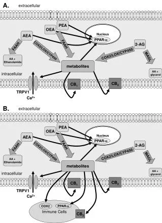
Nucleus
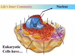
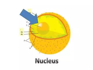
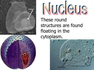
Nucleus. Control center of the cell contains the “genetic library” encoded in the sequences of nucleotides in molecules of DNA code for the amino acid sequences of all proteins determines which specific proteins are to be made in a particular cell type determines the function of that cell

Nucleus
E N D
Presentation Transcript
Nucleus • Control center of the cell • contains the “genetic library” encoded in the sequences of nucleotides in molecules of DNA • code for the amino acid sequences of all proteins • determines which specific proteins are to be made in a particular cell type • determines the function of that cell • The synthesis of proteins involves: • molecules of DNA • enzymes • molecules of RNA • ribosomes
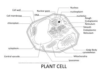
DNA and the Genetic Code • 23 pairs of DNA molecules (46 total) are located in the nucleus of all cells except sperm and oocytes • 23 molecules are inherited from each parent • Recall that DNA is a double stranded molecule of nucleotides that are held together by hydrogen bonds between complimentary bases across the 2 strands • the coding strand and the template strand • T…A and G…C • Each molecule of DNA is subdivided into thousands of segments containing a specific sequence (code) of nucleotides called genes • instruction manual for building proteins • the sequence of nucleotides in the gene’scoding strandcodes for the amino acid sequence of a protein • only the template strand is used for the synthesis of proteins
DNA and the Genetic Code • The alphabet of DNA is A, T, G and C • Within a gene, groups of 3 nucleotides in the templatestrand of DNA form meaningful “words” called triplets • ATG, GCG, TCA, GGT, CAT… (64 different possible combinations) • each triplet codes for a amino acid of the protein encoded by the gene • a gene that is contains 3,000 nucleotides (1,000 triplets) will code for a protein that consists of 1,000 amino acids
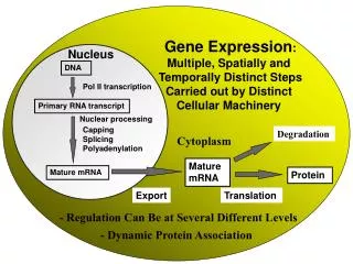
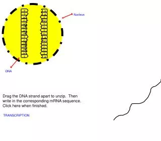
DNA and messenger RNA • Ribosomes, which synthesize all proteins, translate the nucleotide sequence of the DNA strand into the amino acid sequence of a protein • Problem: • the very large molecules of DNA are unable to fit through the nuclear pores to bring the nucleotide code to a ribosome in the cytoplasm • Solution: • an enzyme located in the nucleus called RNApolymerase synthesizes a molecule of single stranded messenger RNA (mRNA) using the template strand of DNA in the nucleus in a process called transcription • mRNA is capable of leaving the nucleus to bring the nucleotide code to a ribosome
Transcription by RNA Polymerase • RNA polymerase • breaks the H-bonds between complimentary nucleotides of DNA strands to separate the coding from the template strand • synthesizes a molecule of mRNA complementary to the template strand of DNA • This synthesizes a molecule of mRNA contains the exact sequence of nucleotides as the coding strand of DNA except for a U for T substitution
mRNA • The alphabet of RNA is A, U, G and C • Within a molecule of mRNA, groups of 3 sequential nucleotides form meaningful “words” called codons • complementary to triplets in the template strand of the gene that was transcribed by RNA polymerase • each codon is a code for an amino acid of the protein coded by the gene • mRNA carries instruction for protein synthesis to a ribosome where it is translated into the primary structure (amino acid sequence) of a protein
Codons • 64 different codons including: • “start” codon (first amino acid of a protein) • always AUG (methionine) • amino acid codons • ACC, GAG, GGG, CAU,… • since there are only 20 amino acids that are used to make proteins, there are multiple codons that code for a single amino acid • “stop” codon (signals the end of the protein) • UAG, UGA, UAA • do NOT code for any amino acid
Translation • Synthesis of a protein molecule by a ribosome • A ribosome “reads” the codons of mRNA from the “start” codon to the “stop” codon • assembles the primary structure of a protein as determined by sequence of codons in mRNA beginning with the start codon and ending with the stop codon • amino acids are brought to the ribosome in the correct order by molecules of transfer RNA (tRNA)
tRNA • Molecules of tRNA are found within the cytosol of a cell which carry amino acids to a ribosome • Each molecule of tRNA: • contains a 3 nucleotide segment on one end of the molecule called the anticodon • complementary to each of the possible codons of mRNA • except for the 3 stop codons • 61 molecules (anticodons) of tRNA • contains a 3 nucleotide segment on the other end of the molecule that attaches to an amino acid
Translation • The codons of mRNA are “read” by a ribosome • When the ribosome reads the start codon, the first amino acid is carried to the ribosome by the tRNA with the complimentary anticodon • the ribosome removes the amino acid from the tRNA • When the ribosome reads the second codon, the second amino acid is carried to the ribosome by the tRNA with the complimentary anticodon • The ribosome removes the amino acid from the tRNA and creates a bond (peptide) between the first and second amino acid • This process continues until the ribosome reads a “stop” codon • no corresponding anticodon • finished protein is “released” from the ribosome
Cell Cycle • The sequence of events in the life of a cell is referred to as the cell cycle • Cell cycles can be long (decades) • nerve cells, muscle cells, fat cells • Cell cycles can be short (few days) • skin cells, stem cells, gastric (stomach) cells • Cells with short cycles can renew themselves through a process called mitosis (cell division)
Cell Cycle • Interphase • the cell is active and provides function in the body • most of the cell cycle is spent in this stage • Mitotic phase • one old cell divides in half to make 2 new cells • short stage compared to interphase
Interphase • First gap phase (G1) • growth • cell size increases • cell contributes structurally and functionally to the organism • Synthesis (S) • preparation for mitosis • DNA replication occurs • each molecule DNA is copied by the enzyme DNApolymerasebefore the cell divides so that each new cell contains the 46 molecules of DNA necessary for normal cellular functioning • Second gap phase (G2) • growth • cell size increases in preparation for cell division • synthesis of enzymes required for mitosis
DNA Replication • 2 DNA Polymerase enzymes are required to replicate a single molecule of DNA • Each DNA Polymerase • unwinds the helical DNA molecule • breaks the H-bonds between the complimentary strands of DNA creating a replication fork • “reads” the sequence of nucleotides along one of the “original” strands of DNA • synthesizes a “new” complementary strand of DNA for each of the “original” strands from free nucleotides in the nucleus • After replication is completed, the cell contains 46 pairs of DNA molecules • 92 total
Semiconservative DNA Replication • The replication of DNA in this manner is considered to be semiconservative because the resulting 2 molecules of double stranded DNA contain one “original” strand and one “new” strand
Mitosis • Process by which one cell divides into 2 identical daughter cells • Functions of mitosis • growth • replacement of old and dead cells • repair of injured cells • Phases of mitosis • Prophase • Metaphase • Anaphase • Telophase
Prophase • Replicated DNA molecules begin to condense into structures that are visible using a compound light microscope called chromatids • 2 genetically identical chromatid pairs are joined together at the centromere by proteins called kinetochores • Nuclear envelope disintegrates releasing the chromosomes into the cytoplasm • Organelles called centrioles move towards opposite sides (poles) of the cell and synthesize mitotic spindle fibers • the mitotic spindle is a web of fibrous proteins called microtubules which are responsible for the equal division of all cellular material between the 2 daughter cells
Metaphase • Metaphase = middle • Spindle fibers from each centriole attach to the kinetochores of the chromatid pairs • allign chromatid pairs to the middle (equator) of the cell • Spindle fibers called asters from each centriole attach to the plasma membrane toanchor centrioles in place
Anaphase • Each centriole retracts the microtubules which pull the sister chromatids away from each other and toward opposite poles of cells • the centromeres split and the 2 chromatids separate • This stage ensures that when the cell divides down the equator, each daughter cell will have 46 molecules of DNA
Telophase • Chromatids extend (loosen) • 2 nuclear envelopes are created around the chromatin • Mitotic spindle breaks down
Cytokinesis • Cytokinesis = cytoplasmmovement • Cytokinesis is the division of the cytoplasm (organelles and intracellular fluid) between 2 newly forming cells • Follows telophase • Creates a crease around cell equator called cleavage furrow • pinches the cell in two