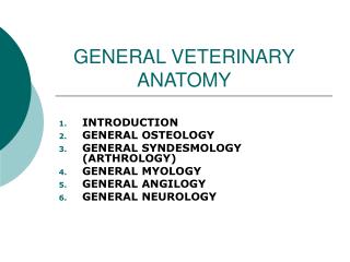
GENERAL VETERINARY ANATOMY
GENERAL VETERINARY ANATOMY. INTRODUCTION GENERAL OSTEOLOGY GENERAL SYNDESMOLOGY (ARTHROLOGY) GENERAL MYOLOGY GENERAL ANGILOGY GENERAL NEUROLOGY. INTRODUCTION.

GENERAL VETERINARY ANATOMY
E N D
Presentation Transcript
GENERAL VETERINARY ANATOMY INTRODUCTION GENERAL OSTEOLOGY GENERAL SYNDESMOLOGY(ARTHROLOGY) GENERAL MYOLOGY GENERAL ANGILOGY GENERAL NEUROLOGY
INTRODUCTION ANATOMY IS THE BRANCH OF BIOLOGICAL SCIENCE WHICH DEALS WITH THE FORM AND STRUCTURE OF ORGANISMS. CLOSE CORELATION WITH PHYSIOLOGY, WHICH TREATS OF THE FUNCTIONS OF THE BODY.
NATURE SCIENCES *ABIOLOGI (physis, mathematic) *BIOLOGI : $ fisiology $ morphology: .anatomy phytotomy : plants zootomy : embryology hystology
ANATOMI • ANATEM (YUNANI): cutting apart or dissociation of part of the body • ANATOMY in the earlier phase of development anatomy was necessarily a purely descriptive science, based on such observation as were possible with unaided eyes and simple dissecting instruments – scalpel, forceps, and the like.
GENERAL ANATOMY • MACROSCOPIC ANATOMY ( GROSS ANATOMY) • MICROSCOPIC ANATOMY ( HISTOLOGY = TISSUE SCIENCE)
THE DEVELOPMENT & GROWTH OF THE ORGANISM • EMBRYOLOGY : the earlier phase of development of organism during which the tissue and organ are formed: fertilisation – foetus - partus • ONTOGENY: study of the development of the individual organism • FILOGENY (ancestral history) : is constituted by the evolutionary changes which it has undergone, as disclosed by geological record.
ORGANISM AS THE OBJECT OF THE STUDY *SPECIAL ANATOMY : is the description of the structure of a single or species : anthropotomy, kinotomy dan hippotomy * COMPARATIVE ANATOMY :is the description and comparison of the structure of animals
HOW TO STUDY ANATOMY? • SYSTEMATIC ANATOMY • TOPOGRAPHIC ANATOMY • APPLIED ANATOMY
SYSTEMATIC ANATOMY • Osteology • Syndesmology • Myology • Splanchnology • Angiology • Neurology • Aesthesiology • Sense organs • Common integument
NOMENCLATURE (NAV, 1972) • Until 1895 there was no general agreement on the nomenclature of human or veterinary anatomy. Each nation had its own systemof terminology. • The first international Nomina Anatomica Veterinaria was published in 1968.
AGREEMENT IN THE N.A.V • 1. Aside from a very limited number of exception, each anatomical concept should be designated by a single term. • Each term should be in Latin in the official list, but the anatomists of each country are free to translate the official Latin terms into the language of instruction.
3. each term should be as short and simple as possible. • 4. The term should be easy to remember and should have, above all, instructive and discriptive value. • 5. Structure that are closely related topographically should have similar names; exp: arteria femoralis, vena femoralis, nervus femoralis.
6. Differentiating adjectives should generally be opposites, as major and minor, superficial and profundus. • 7. Term derived from proper names (eponyms) should not be used.
DESCRIPTIVE TERM IN LATIN THIS TERM USE IN THE BODY • Dorsal • Ventral • Cranial • Caudal • anal dorsal cranial caudal ventral
The terms cranialis and caudalis apply to to the neck, trunk, tail, and the limb as far distally as the end of the antebrachium and crus. • The terms dorsalis and palmaris are used on the manus, and dorsalis and plantaris on the pes. • On the head the term rostralis, caudalis, dorsalis and ventralis are preferred, with terms anterior, posterior, superior, and inferior used in few location, such as eye ball, eyelids,and inner ear.
Medialis and lateralis are used on the whole body, except that axialis and abaxialis designate the sides of the digits in domestics mammals other than the horse.
Proximal - distal Proximal: the direction go up Distal : the direction go down
Cranial/rostral - aboral aboral rostral oral
ANATOMICAL DESCRIPTION dorsal cranial anal caudal CRANIAL ventral
Oral Apical Aboral Nuchal Anterior posterior Superior Inferior This term only in the skull
Proximal Distal Dorsal Volar Palmar plantar Ulnar Radial fibular Tibial Lateral Medial Median Sagittal Transversal horizontal Extremities (thoracic and pelvic limb)
Plana median Median: devided the body in the median plane bilateral simetrical.
SAGITTAL Cutting the body in the paralel ways with the median plane.
Dexter Sinister Externus Internus Profundus Superficialis Transversus longitudinal Ecto Meso Endo Epi Peri Dia Hypo dan Hyper basis dan apex margo THE DIRECTION ORIENTATION
CONDITION OF THE BONE • Magnus • Brevis • Major/ majus • Minor/ minus • Dorum • Molle • Supra dan infra
facies Fovea Facialis Fascia Foramen Sulcus Fasciculus canalis Cavum Caverna(cavernosus) Caput Condylus Collum Spina Crista incissura MORPHOLOGY OF BONE
PROC TRANSVERSUS Proc transversus
PROC SPINOSUS Proc spinosus
RADIOLOGY ANATOMY • X RAY was invented in 1895 by CONRAD ROENTGEN. • It useful for diagnosis, treatments and research. • To be proficient in radiographic interpretation, one must first have some knowledge of the anatomy of the region x-rayed.
Three elements in x-ray shadow • 1.AIR : in mouth, nose, paranasal sinuses, trachea, lungs, stomach, small intestine of the nursing individual, colon, and rectum. • 2. WATER: in the blood (large vessels) and blood filled organs, such as liver, spleen and kidneys, and the urine-filled bladder. • 3. MINERALS:primary significance, the calcium found in bones. Bone has calcium content & high radiopacity
Developmental anatomy • FERTILISASI ZYGOTE (mitosis: cleavage) MORULA BLASTULA GASTRULA devide into 3: - ectoderm : external layerskin & nerve - endoderm: internal layer viscera - mesoderm: cells between external and internal layers muscles&bone
THE FUNCTION OF SKELETON • To support the body • To give the body form • To protect the soft and weak tissue • As a passive locomotion • As a place for attachment of the muscles • To produce blood. • As chemical reservoir agents: Ca & P
THE AMOUNT of THE BONES • Every breed have different amount • examples: horse 205 bones catlle 191 – 193 bones chicken 160 bones human 206 bones (old), 270 ( baby) This condition is depend on: breed & age
POSITION of the SKELETON • Axial skeleton • Appendix skeleton • Visceral skeleton : example: os penis : dog & cat os cordis : sapi os glandis : cat os hyodeus: vertebrata
ACCORDING TO THE SHAPE OF THE BONES • OSSA LONGA • OSSA PLANA • OSSA BREVIA • OSSA IRREGULARIA
DEVELOPMENT OF THE BONES(osteogenesis) • 1.osteogenesis intramembranosa (desmalis = primer): mesenchym cells osteoblast osteocyt matrix become jelly & solid (osteoid) calcification punctum ossification. • 2. osteogenesis intracartilagenosa (enchondralis = secundair): it begin with cartilago: mesenchym cells chondroblast chondrocyt (fill according the length of the bone) ossification. Osteoblast : cells that destroy the bone cells which already done in order to have good shape.
ossification center DEVELOPMENT : • interstitial development ( from the middle of the tissue) • appostitional development (from the lateral, usually from perichondrium or periosteum become mass bone)
group I horse none cattle none sheep none group II human 31 rabbit 32 dog 34 cat 34 pig 3 Guinea pig 3 PUNCTUM OSSIFICATION VERTEBRATA post natal
Maturity • 1. sex maturity : genital organ already have their function properly. ♂: wet dreams & ♀: menstruation • 2. body maturity : all the bones already done (don’t have any punctum ossification ossification has alredy finished means the growth stop.
Sexual & body maturity Breed sex mature body mature • horse 1 thn 4-5 thn • cattle 5-9 bln 4-5 thn • Sheep/goat 6 bln 4-5 thn • pig 3-4 bln 4-7 thn • dog 8 bln 1,5-2 thn
Structure of the bones • macroscopic structure • microscopic structure • physical and chemical structure
MACROSCOPIC STRUCTURE SUBSTANSIA SPONGIOSA SUBSTANSIA COMPACTA
Mass and spons bones os spongiosa os compact
long bone EPIPHYSA DIAPHISA