Download
1 / 1
10 likes | 167 Vues
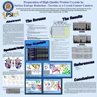
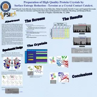
Salt Screen. Standard Screen. #. #. Hits. Hits. Precipitant. Precipitant. Salt. Salt. Buffer. Buffer. Like. Like. 73. 75. 11. 8. 30%(w/v) PEG 8000. 30%(w/v) PEG 4000. 0.2 M (NH 4 ) 2 SO 4. 0.2 M Li 2 SO 4. 0.1 M Cacodylate pH 6.5. 0.1 M Tris pH 8.5. H1 #15. H1 #17. 75.

E N D
Salt Screen Standard Screen # # Hits Hits Precipitant Precipitant Salt Salt Buffer Buffer Like Like 73 75 11 8 30%(w/v) PEG 8000 30%(w/v) PEG 4000 0.2 M (NH4)2SO4 0.2 M Li2SO4 0.1 M Cacodylate pH 6.5 0.1 M Tris pH 8.5 H1 #15 H1 #17 75 41 6 7 30%(w/v) PEG 4000 30%(w/v) PEG 8000 0.2 M Li2SO4 0.2 M Li2SO4 0.1 M Tris pH 8.5 0.1 M NaAcetate pH 4.5 H1 #17 W1 #17 85 76 6 5 2.0 M (NH4)2SO4 20%(w/v) PEG 8000 0.2 M MgAcetate 0.1 M Cacodylate pH 6.5 H1 #32 H1 #18 70 89 5 5 1.4 M Na Citrate 30%(w/v) PEG 4000 0.2 M (NH4)2SO4 0.1 M HEPES pH 7.5 0.1 M NaCitrate pH 5.6 H1 #38 H2 #9 43 70 5 4 20%(w/v) PEG 8000 30%(w/v) PEG 4000 0.2 M (NH4)2SO4 0.2 M MgCl2 0.1 M NaCitrate pH 5.6 0.1 M Tris pH 8.5 H2 #9 W2 #2 2 5 20%(w/v) PEG 3000 0.1 M NaCitrate pH 5.5 W1 #6 Distribution of Hits Standard Screen Target Residue Ser Thr His Tyr Standard Salt Standard Salt Overlaps Standard Salt Overlaps Standard Salt Overlaps Standard Salt Overlaps Total Unique Total Unique Mutant Distribution of Hits Salt Screen 0.1M bicine pH=7.5 30% PEG 6000 32% PEG 8000, 0.22M (NH4)2SO4, 0.1M cacodylate pH=6.5 Resolution 2.1Å Spacegroup P21 (a=32.0,b=55.1,c=38.9,=107.5) Discontinued: Solubility Problems A B C D Old and New RhoGDI Crystal Forms E F G H I Previous Xtals New Xtals Preparation of High Quality Protein Crystals by Surface Entropy Reduction - Tyrosine as a Crystal Contact Catalyst. Tomek Boczek, Gosia Sikorska, Kasia Grelewska, Gosia Pinkowska, Michal Zawadzki, David R. Cooper and Zygmunt DerewendaPSI Center for Structure and Function Innovation and Department of Molecular Physiology and Biological Physics University of Virginia. Charlottesville, VA, 22908 The Results The Screens Abstract Crystallization is a limiting step in macromolecular crystallography. In cases where proteins are particularly recalcitrant to crystallization efforts, mutational modification of surface properties may be essential. We previously suggested that targeted replacement of clusters of residues with high-conformational entropy (lysines, glutamates and/or glutamines) with alanines leads to formation of epitopes that are capable of mediating crystal contacts. This is because the entropic cost of immobilizing large side chains at the intermolecular contact regions has been reduced and crystal contacts can be formed by the mutated epitopes. This Surface Entropy Reduction (SER) method has facilitated the crystallization and structure determination of a number of novel proteins and has also led to the discovery of crystal forms that diffract to significantly higher resolution than the wild-type form. However, it has not been conclusively demonstrated if alanine constitutes the best choice for replacement of high-entropy residues. Here we present a systematic study of the replacement of nine Lys/Glu-rich patches in RhoGDI with four target residues; Ser, Thr, His and Tyr. All four amino acids are known to occur at interfaces with significantly higher incidence than Lys or Glu / Gln, and may mediate weak protein-protein interactions leading to crystal formation. Our results show that tyrosine is a particularly good choice for the target amino acid, with threonines also performing quite well. The mutated residues often participate in crystal contacts, in both homotypic (symmetric) or heterotypic (head-to-tail) intermolecular interactions. We also examined a crystallization method proposed by Janet Newman that replaces the normal crystallization reservoir solutions with 1.5 M NaCl. The results are very promising; with more than half of the mutants in this series yielding more crystals when salt was used as the reservoir solution. Moreover, this method greatly increased the variety of conditions that yielded crystals, with little overlap of the conditions that yielded crystals for the two types of screens. This suggests a crystallization strategy for proteins for which crystallization is the major bottleneck. Creating several mutants by replacing patches glutamates and lysines with two or more target residues and conducting screens with normal and alternate reservoir solutions greatly increases the chances of obtaining diffraction quality crystals • The Super Screen • We use a custom 96 well screen we had Nextal generate for us. • For details see, • http://ginsberg.med.virginia.edu/~dcoop/Superscreen • We use sitting drop setups. For all of our screens, the protein was concentrated to 15 mg/ml. • Standard Screens • Drops are mixed 1:1 with protein solution and Super Screen reagent. • Reservoir is 100 l of Super Screen reagent. • Salt Screens • As above, drops are mixed 1:1 with protein solution and Super Screen reagent. • Reservoir is 100 l of 1.5 M NaCl The Crystals Experimental Design Model Protein : RhoGDI (66-204) RhoGDI posses all of the characteristics of proteins that lend themselves to crystallization by the SER method. It expresses and purifies well and can be concentrated easily, but wild-type RhoGDI is difficult to crystallize. Additionally RhoGDI is rich in lysines (10.1% -- average frequency is 7.2% ) and glutamates (7.9% -- average frequency is 3.7%), giving us many potential mutation sites. The Mutant Series Nine mutants were chosen, with the mutations designed to reduce or eliminate clusters of high-entropy residues. The mutants were designated by two letters; the first letter indicates which mutations the mutant contains and the second letter designates the target amino acid. Thus, the CY mutant is K135Y, K138Y, K141Y. The B mutant was discontinued do to low expression or solubility. Conclusions All RhoGDI mutants were expressed as a GST-fusion protein. After cleavage of the fusion protein with rTEV, GST was removed by size exclusion chromatography. GST-RhoGDI RhoGDI Initial Purification Size Exclusion Purification