Download
1 / 34
460 likes | 1.98k Vues
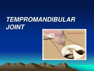
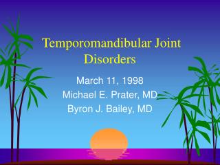
By: Nour-Eldin A. Nour-Eldin Mohammed. The Tempromandibular Joint (TMJ). Normal Anatomy. Mandibular condyle (head). Glenoid fossa. Articular tubercle (eminence). Posterior band of articular disc. Anterior band of articular disc. Mandibular condyle (head).

E N D
By: Nour-Eldin A. Nour-Eldin Mohammed The Tempromandibular Joint(TMJ)
Normal Anatomy Mandibularcondyle (head) Glenoidfossa Articular tubercle (eminence)
Posterior band of articular disc Anterior band of articular disc Mandibular condyle (head) Lateral pterygoid muscle raphe Lower head of lateral pterygoid muscle Posterior disc attachment
Mandibular condyle (head) Articular disc
MRI and autopsy sections: upper row oblique sagittal MRI, asymptomatic volunteer: left lateral, middle medial, right opened mouth
Internal Derangements • General orthopedic term implying a mechanical fault that interferes with the smooth action of a joint • The most common internal derangement is disc displacement Clinical Features • Clicking sounds from joint(s) • Restricted or normal mouth opening capacity • Deviation on opening • Pain
Internal Derangements Imaging Features • Anterior disc displacement: posterior band of the disc located anterior to the superior portion of the condyle at closed mouth on oblique sagittal images • Disc may have normal (biconcave) or deformed morphology • In opened mouth position disc may be in a normal position (“with reduction”) or continue to be displaced (“without reduction”)
Partial anterior disc displacement at baseline lateral sections central sections open-mouth
Complete anterior disc displacement medial section Autopsy Open-mouth MRI
Medial disc displacement coronal MRI Oblique coronal MRI
Osteoarthritis Definition • Non-inflammatory focal degenerative disorder of synovial joints, primarily affecting articular cartilage and sub-condylar bone; initiated by deterioration of articular soft-tissue cover and exposure of bone. Clinical Features • Crepitation sounds from joint(s) • Restricted or normal mouth opening capacity • Pain or no pain from joint areas and/or of mastication muscles • Occasionally, joints may show inflammatory signs • Women more frequent than men
anteriorly displaced and deformed, degenerated disc and irregular cortical outline with osteophytosis and sclerosis of condyle .
Advanced osteoarthritis and anterior disc displacement, with joint effusion
Bone Marrow Abnormalities Definition • Bone marrow edema: serum proteins within marrow interstitium surrounded by normal hematopoietic marrow. • Osteonecrosis: complete loss of hematopoietic marrow.
Imaging Features • Abnormal signal on T2-weighted image from • condyle marrow: increased signal indicates marrow edema; reduced signal indicates marrow sclerosis or fibrosis • Combination of marrow edema signal and marrow sclerosis signal in condyle most reliable sign for histologic diagnosis of osteonecrosis • Marrow sclerosis signal may indicate advanced • osteoarthritis without osteonecrosis, or osteonecrosis
Arthritides Definition • Inflammation of synovial membrane characterized by edema, cellular accumulation, and synovial proliferation (villous formation). Clinical Features • Swelling of joint area, not frequently seen in TMJ • Pain (in active disease) from joints • Restricted mouth opening capacity • Morning stiffness, in particular stiff neck • Dental occlusion problems; “my bite doesn’t fit” • Crepitation due to secondary osteoarthritis
Rheumatoid arthritis. After 1 year
Rheumatoid arthritis. A MRI shows completely destroyed disc, replaced by fibrous or vascular pannus and cortical punched-out erosion (arrow) with sclerosis in condyle.
Psoriatic arthropathy. Obliquecoronal and oblique sagittal CT images show punched-out erosion in lateral part of condyle (arrow). Psoriatic arthropathy. MRI shows contrast enhancement within bone erosion and in joint space, consistent with thickened synovium/pannus formation. Openmouth MRI shows reduced condylar translation but normally located disc (and normal bone in this section)
Ankyloses Definition Fibrous or bony union between joint components.
Growth Disturbances (Anomalies) Definition Abnormal growth of mandibular condyle; overgrowth, undergrowth, or bifid appearance.
Condylar hypoplasia and facial asymmetry Condylar Hypoplasia Normal TMJ
Inflammatory or Tumor-like Conditions Calcium Pyrophosphate Dehydrate Crystal Deposition Disease (Pseudogout)
Benign Tumors Synovial Chondromatosis • Benign tumor characterized by cartilaginous metaplasia of synovial membrane, usually in knee, producing small nodules of cartilage, which essentially separate from membrane to become loose bodies that may ossify.
Osteochondroma Definition Benign tumor characterized by normal bone and cartilage, near growth zones.
Malignant Tumors Osteosarcoma mandible; 18-yearold female
Malignant tumor, mandible; 70- year-old male with metastasis from lung cancer


![TEMPROMANDIBULAR JOINT AND MOVEMENTS MANDIBULAR [ T M J ]](https://cdn3.slideserve.com/6414015/slide1-dt.jpg)